Case 56

 Clinical Presentation
A 63-year-old woman with flank pain for 2 days. There is no history of trauma.
 Imaging Findings

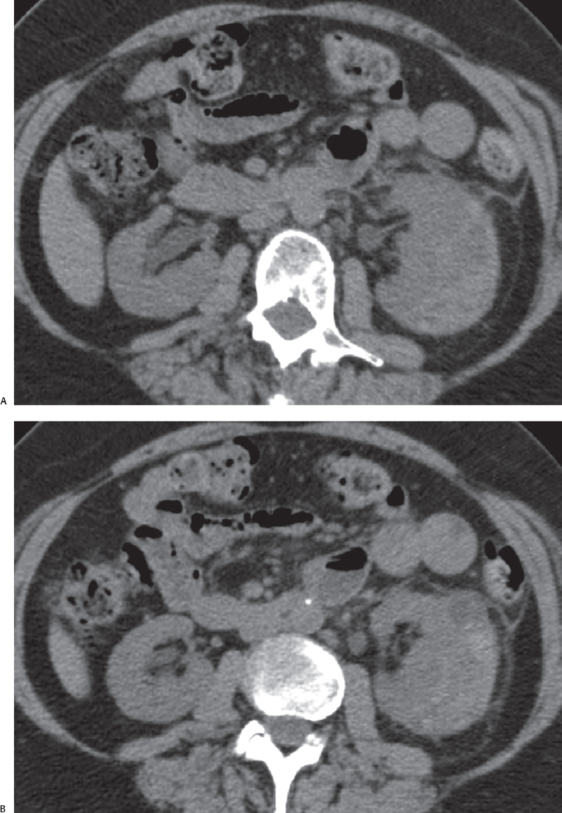
(A) Precontrast computed tomography (CT) image of the abdomen in the region of the kidneys shows a high-attenuation collection (arrow) at the lateral aspect of the left kidney. The attenuation of the collection is suggestive of blood. Significant perinephric stranding is seen (arrowhead). (B) Precontrast CT image of the abdomen in the region of the kidneys at a level lower than that in Figure A again shows the high-attenuation collection (arrow). However, there is a small focal lesion in the renal parenchyma at this level (arrowhead). (C) Contrast-enhanced CT image of the abdomen at a level comparable to that in Figure B obtained 6 weeks later shows marked enhancement of the focal lesion seen in Figure B (arrowhead). No fat is seen. The previously seen collection appears to have more fluid attenuation and is smaller (arrow).
 Differential Diagnosis
• Acute subcapsular hematoma from rupture of a renal cell carcinoma:
Stay updated, free articles. Join our Telegram channel

Full access? Get Clinical Tree


