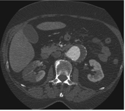
CASE 56 A patient with a known abdominal aortic aneurysm and recent cardiac catheterization presents with new-onset right flank pain. Fig. 56.1 Contrast-enhanced axial CT image shows a lack of enhancement in a geographic wedge-shaped area of the right kidney. The left kidney enhances normally. An infrarenal aortic aneurysm is also seen. Contrast-enhanced axial computed tomography (CT) image shows a lack of enhancement in a geographic wedge-shaped area of the right kidney (Fig. 56.1). The left kidney enhances normally. An infrarenal aortic aneurysm is also seen. Renal infarct following angiographic catheter manipulation in the setting of an abdominal aortic aneurysm Renal infarction has a variety of causes. It most commonly occurs in patients with a history of thromboembolic disease.
Clinical Presentation
Radiologic Findings
Diagnosis
Differential Diagnosis
Discussion
Background
Clinical Findings
Related posts:


Stay updated, free articles. Join our Telegram channel

Full access? Get Clinical Tree


