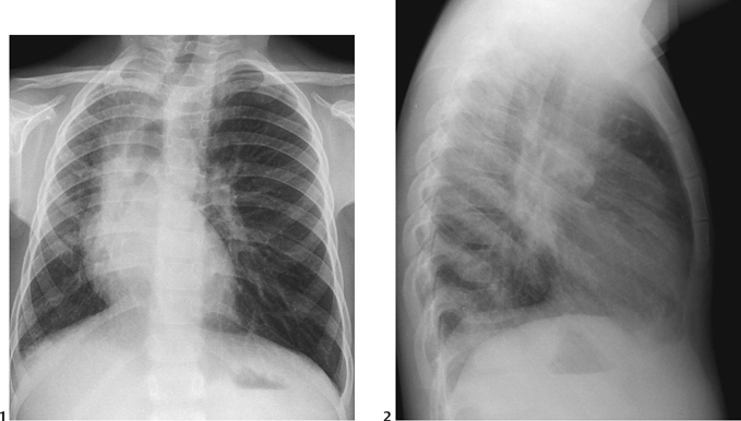
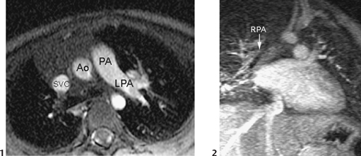
CASE 56 A 5-year-old child presents with recurrent pneumonia. Figure 56A A frontal chest radiograph (Fig. 56A1) shows situs solitus and levocardia. The heart is positioned on the right side with small right lung volume. The pulmonary vascularity is asymmetric with prominent vessels in the left lung. The right lung shows reticular pattern. The lateral view (Fig. 56A2) is not remarkable. Figure 56B Bright-blood MRI in axial plane (1) shows that right pulmonary artery is missing. Ao, aorta; LPA, left pulmonary artery; PA, main pulmonary artery; SVC, superior vena cava. MR angiogram in an oblique coronal view (2) of the same patient shows tiny right pulmonary artery (RPA) in the right lung hilum. Unilateral absence of the right pulmonary artery. An axial bright-blood MRI (Fig. 56B1) shows absence of the mediastinal segment of the right pulmonary artery. A contrast-enhanced MR angiogram (Fig. 56B2) shows the tiny right pulmonary artery and its branches in the right lung. Hypoplastic right lung The clinical presentation of congenital unilateral absence of a pulmonary artery may be subtle. Patients often have a history of recurrent pulmonary infections. Hemoptysis from dilated bronchial arteries may develop in older children and adults. Once one is familiar with the diagnosis, it may be suspected from a chest radiograph.
Clinical Presentation
Radiologic Findings
Diagnosis
Differential Diagnosis
Discussion
Clinical Findings
Pathology
Related posts:


Stay updated, free articles. Join our Telegram channel

Full access? Get Clinical Tree


