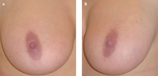
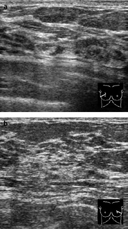
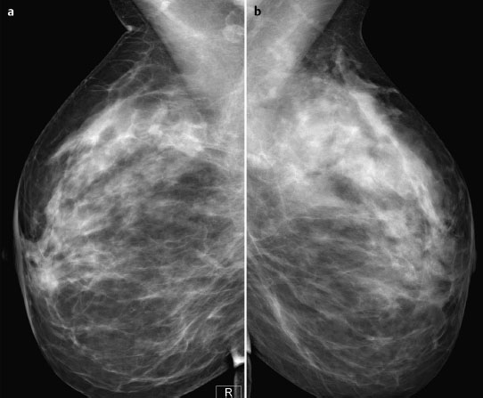
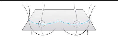
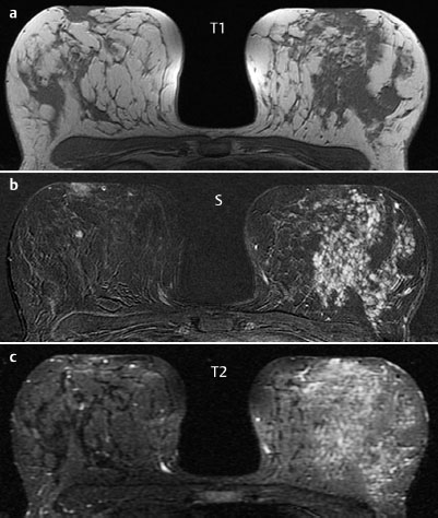
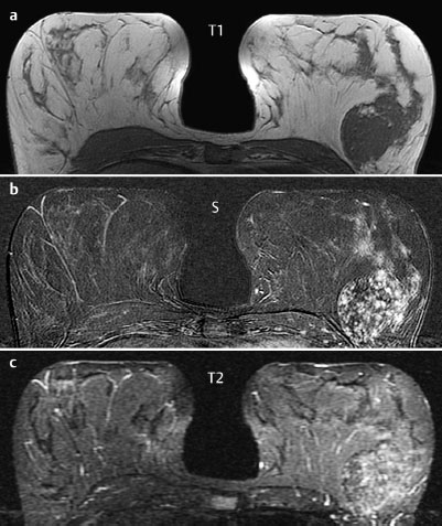
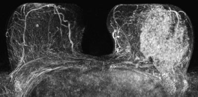
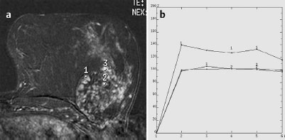
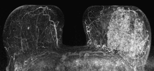
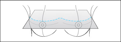
Case 57 Indication: Mastodynia of the left breast. History: Unremarkable. Risk profile: No increased risk. Age: 43 years. Fig. 57.1 a,b Clinical examination of the right (a) and left (b) breasts. Fig. 57.2a,b Sonography. Fig. 57.3a,b Digital mammography, MLO view. Fig. 57.4a–c Contrast-enhanced MRI of the breasts. Fig. 57.5a–c Contrast-enhanced MRI of the breasts. Fig. 57.6 Contrast-enhanced MR mammography. Maximum intensity Fig. 57.7a,b Signal-to-time curves. projection. Please characterize ultrasound, mammography, and MRI findings. What is your preliminary diagnosis? What are your next steps? The imaging of a young woman with unilateral mastodynia is presented. The clinical aspect of both breasts was normal. No resistance in the area of the pain in the left breast. Ultrasound demonstrated a marked difference between the echo textures of left and right breasts. There were no other abnormalities and no signs of malignancy. US BI-RADS right 1/left 1. Mammography showed asymmetry of the parenchyma (right breast was inhomogenously dense, ACR type 3, left partially extremely dense, ACR type 4). No unusual findings. No densities or architectural distortion. No calcifications (BI-RADS right 1/left 2). PGMI: G (inframammary fold not correctly positioned). As expected, the precontrast MRI showed asymmetry of the parenchymal structure (left>right). In the outer upper quadrant of the left breast there was a circumscribed, well-defined section of parenchyma. After contrast administration there was a marked difference between the enhancement pattern in each breast: the right breast demonstrated only a small, adenoma-like focal enhancement, whereas the left breast showed pronounced patchy enhancement throughout the parenchymal tissue as well as within the capsule-like circumscribed formation. MRI Artifact Category: 2 MRI Density Type: 1

Examination
Ultrasound
Mammography
MR Mammography
MRM score | Finding | Points |
Shape | round | 0 |
Border | well-defined | 0 |
CM Distribution | homogenous | 0 |
Initial Signal Intensity Increase | strong | 2 |
Post-initial Signal Intensity Character | wash-out | 2 |
MRI score (points) |
| 4 |
MRI BI-RADS |
| 4 |
 Differential Diagnosis
Right: Normal tissue with adenoma.
Left: Hamartoma (capsule-like area), adenosis, diffuse carcinoma.
Clinical Findings | right 1 | left 1 |
Ultrasound | right 1 | left 1 |
Mammography | right 1 | left 2 |
MR Mammography | right 2 | left 4 |
BI-RADS Total | right 2 | left 4 |
Procedure
Histological investigation of the diffuse patchy enhancement seen in the left breast in MRI. Considering the homogeneity of the enhancement pattern, MR-guided vacuum biopsy was not required. Instead a US-guided representative “blind” biopsy was performed in three different regions of the left breast (upper outer quadrant, lateral to the nipple, and lower outer quadrant).
Histology of the left breast
Mastopathy. Additionally, in one of the specimens a circumscribed region containing part of a “carcinoma lobulare in situ” was found. This carcinoma had minimal proliferative activity. There was no atypical ductal proliferation and no invasive lobular carcinoma. There was no suggestion of a ductal carcinoma in situ or an invasive carcinoma. These histology results were confirmed by supplementary immunohistochemical analysis.
Further Procedure
A four-week trial of antihormonal therapy followed by repeat MR mammography. This repeat imaging showed no change in the findings of unilateral diffuse enhancement of the left breast (Fig. 57.8).
Fig. 57.8 MR mammography with largely unchanged findings following four weeks of anti-hormonal therapy.
Further procedure
Although the histology revealed a CLIS in the core biopsy specimen, an open biopsy was avoided, because the blind biopsy results meant that a very extensive section of tissue from the outer quadrants of the left breast would be excised. In view of the unchanged enhancement of the left breast in MRI after 4 weeks of antihormonal therapy, close monitoring after a further 3, 6 and 12 months was recommended.
Stay updated, free articles. Join our Telegram channel

Full access? Get Clinical Tree


