Case 57
 Clinical Presentation
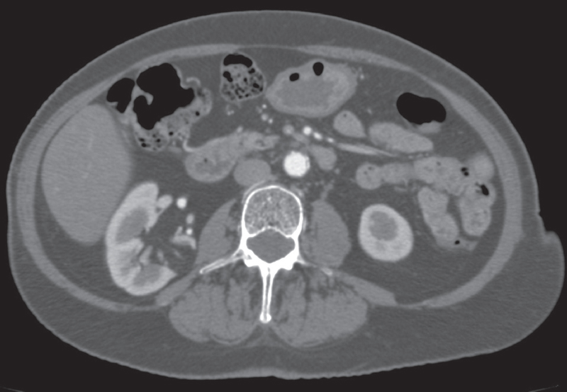
A 39-year-old woman who underwent computed tomography for vague abdominal symptoms.
 Imaging Findings
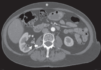
Contrast-enhanced computed tomography (CT) image at the level the kidneys shows a 2.5-cm–diameter focal lesion (arrow) with attenuation similar to that of the subcutaneous fat (asterisk). The lesion extends from the renal sinus to the renal capsule. A vessel (arrowhead) is seen coursing in the mass. No calcifications are noted.
 Differential Diagnosis
• Angiomyolipoma: With very few exceptions, a fat-containing lesion in the renal parenchyma is an angiomyolipoma. Vascularity and soft tissue within the tumor are an inherent part of the neoplasm.
• Fat-containing renal cell carcinoma: Renal cell carcinoma may incorporate fat by various mechanisms. However, this is extremely rare. Other than in a few recent case reports, almost all fat-containing renal cell carcinomas also show calcifications.
• Retroperitoneal liposarcoma:
Stay updated, free articles. Join our Telegram channel

Full access? Get Clinical Tree


