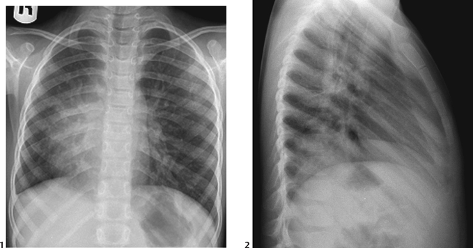
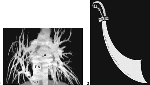
CASE 57 A 5-year-old child presents with recurrent pneumonia. Figure 57A A frontal chest radiograph (Fig. 57A1) shows situs solitus. The heart is positioned on the right with small right lung volume. The right mediastinal border is indistinct. In lateral view (Fig. 57A2), there is a band of increased density behind the sternum. The pulmonary vascularity is asymmetric with prominent vessels noted on the left. Figure 57B CT angiogram reformatted in coronal plane (1) shows a vertical vein (ss) that drains all pulmonary veins from the right lung into the right atrium (RA)-inferior vena cava (IVC) junction. The vertical vein is similar to a scimitar (2), a Turkish sword. The left pulmonary veins connect to the left atrium (LA). Scimitar syndrome. A CT angiogram (Fig. 57B1) shows a scimitar-shaped vein draining the entire right lung to the inferior vena cava at its junction with the right atrium. Hypoplastic right lung The clinical expression of this syndrome is diverse. Some patients present with severe congestive heart failure, whereas others show pulmonary artery hypertension in infancy or early childhood. Not infrequently, scimitar syndrome is recognized in a child with recurrent pulmonary infections. It is often an incidental finding at a routine chest radiographic check in an otherwise asymptomatic adult. Factors contributing to early presentation include severely obstructed pulmonary venous connections, association with other complex cardiac malformations, and/or the presence of a large aberrant systemic arterial supply. A scimitar is a short, curved Turkish sword (Fig. 57B2
Clinical Presentation

Radiologic Findings

Diagnosis
Differential Diagnosis
Discussion
Clinical Findings
Pathology
![]()
Stay updated, free articles. Join our Telegram channel

Full access? Get Clinical Tree


