Case 58
Indication: Monitoring post mastectomy.
History: Breast cancer and mastectomy of the right breast 23 years previously.
Risk profile: Increased by previous incidence of breast cancer.
Age: 73 years.
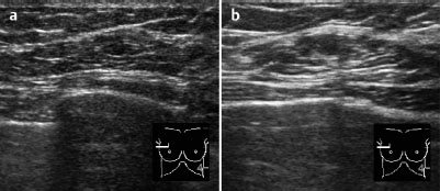
Fig. 58.1 a,b Ultrasound.
Clinical Findings
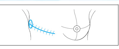
Slight skin thickening in the axillary extension of the mastectomy scar. This was first noted by the patient after a mosquito bite 3 weeks before the examination.
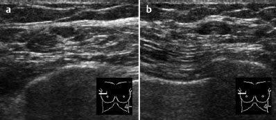
Fig. 58.2a,b Ultrasound.
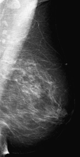
Fig. 58.3 Digital mammography of the left breast, MLO view.
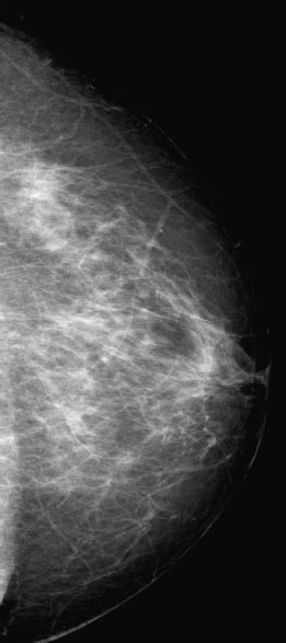
Fig. 58.4 Digital mammography of the left breast, CC view.
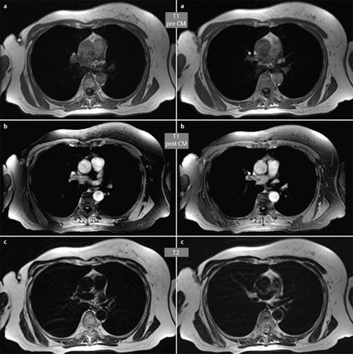
Fig. 58.5,6 MRI of the chest.

|
Please characterize ultrasound, mammography, and MRI findings.
What is your preliminary diagnosis?
What are your next steps?
|
This case demonstrates the follow-up of a woman 23 years after Halsted radical mastectomy for breast cancer. Clinically there is a slight, recently developed thickening in the axillary part of the scar.
Ultrasound
There were no unusual sonographic findings within the scar. In the area around the axillary end of the scar there were two lymph nodes each with a diameter of 6 mm. US BI-RADS 1.
Mammography
Mammograms showed that the parenchyma of the left breast was fibroglandular, ACR type 2. There were no masses, densities, or microcalcifications. After Halsted mastectomy a mammography of the residual breast tissue is not usually possible. BI-RADS left 1.
MR Imaging
After marking of the slight axillary thickening with a nitro capsule, a subcutaneous enhancement was visible in this region. There were no further relevant findings. No additional information was supplied by T2-weighted images.
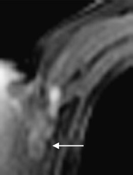
Fig. 58.7 Enlarged post-contrast image of the MRI of the chest.
Analysis of the enhancing area using the Göttingen-Score is not useful in this case.
The general patterns observed following surgery and and healing are:
Scar tissue does not show enhancement post-contrast.
Local tumor recurrence shows enhancement post-contrast.
Caution: Focal inflammation may lead to false positive findings!
MRI BI-RADS 4


