CASE 58
Clinical Presentation
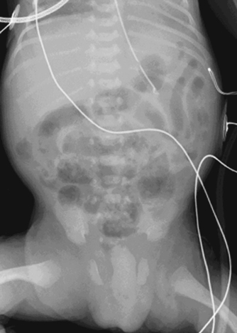
A 6-day-old baby born at 27 weeks of gestation presents with a distended abdomen and hemodynamic instability.
Figure 58A
Radiologic Findings
Supine radiograph (Fig. 58A) demonstrates a bubbly appearance in the lower abdomen consistent with pneumatosis in association with increased interloop distance in the left upper quadrant secondary to bowel wall thickening.
Necrotizing enterocolitis (NEC)
Differential Diagnosis
- Intraluminal fecal residue may resemble mottled appearance of pneumatosis but rarely present in the first 2 weeks of life.
- Focal intestinal perforation without NEC and idiopathic gastric perforation present with pneumoperitoneum but without evidence of pneumatosis
- Mesenteric thromboembolism (catheter-related)
- Hirschsprung’s enterocolitis (term infant)
- Malrotation and volvulus
- Other causes of neonatal sepsis may mimic clinical features of NEC and cause generalized bowel dilatation.
Discussion
Background
NEC is a severe inflammatory enteritis occurring in up to 35% of premature infants, the majority <2000 g. Any part of the bowel may be affected from the esophagus to the rectum, although the distal ileum and ascending colon are most frequently involved.
Etiology
- Multiple etiologic factors have been implicated, including hypoxia, sepsis, hypotension, intrauterine growth retardation, exchange transfusion, and umbilical arterial catheterization. Case clusters suggest an infectious agent is involved, but this may be a secondary event following compromise of the intestinal mucosal barrier. Early introduction of enteral nutrition (particularly formula feeds) and rapid increases in feed volume are associated with a higher incidence. Bowel ischemia appears to be the common pathway, with proliferation of the intestinal flora leading to bowel necrosis and perforation.
- Experimental evidence suggests that the balance of inflammatory mediators, including tumor necrosis factor, platelet activating factor, nitric oxide, and free radicals, influences intestinal perfusion and the integrity of the mucosal barrier. Disturbance of this balance increases vulnerability to NEC.
- NEC may also occur in term infants. Predisposing factors include congenital heart disease (decreased systemic perfusion) or recent abdominal surgery (particularly gastroschisis or intestinal atresia repair), polycythemia, and maternal cocaine abuse.
Clinical Findings
Symptoms usually occur in the first week of life, with very low birth weight infants (<1000 g) often presenting slightly later, after 2 weeks. Clinical signs are varied and may be nonspecific. Abdominal distension, vomiting, diarrhea, or blood per rectum occurs in association with systemic signs including fever, apnea, metabolic acidosis, and, in advanced cases, circulatory collapse. On examination there may be erythema of the abdominal wall and palpable distended bowel loops.
Complications