CASE 58 Incidental finding in a 26-year-old patient complaining of vague abdominal pain. Fig. 58.1 (A,B) Ultrasound scans reveal a well-circumscribed, heterogeneous mass in the upper pole of the left kidney with a central linear region of decreased echogenicity. (C) Contrast-enhanced axial image demonstrates a solid, heterogeneously enhancing mass in the medial interpolar region of the left kidney with an enhancement pattern similar to normal adjacent renal parenchyma. There is a central, nonenhancing portion within the mass. No fat is seen within the lesion. (D) Axial gradient echo T1-weighted image obtained in the arterial phase after gadolinium injection shows a well-demarcated arterially enhancing lesion with a central scar. Ultrasound examination reveals a well-circumscribed, heterogeneous mass in the upper pole of the left kidney with a central linear region of decreased echogenicity (Fig. 58.1A,B). The contrast-enhanced axial image demonstrates a solid, heterogeneously enhancing mass in the medial interpolar region of the left kidney with an enhancement pattern similar to normal adjacent renal parenchyma. There is a central, nonenhancing portion within the mass. No fat is seen within the lesion (Fig. 58.1C
Clinical Presentation
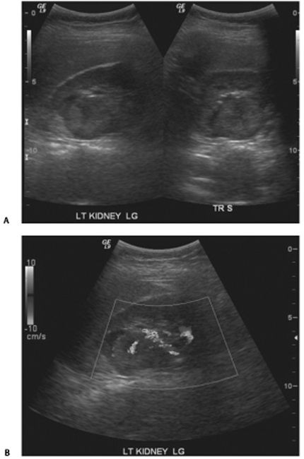
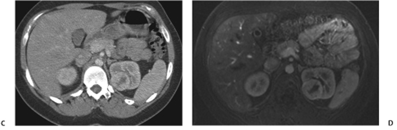
Radiologic Findings
![]()
Stay updated, free articles. Join our Telegram channel


Full access? Get Clinical Tree


