Case 59
 Clinical Presentation
A 48-year-old man with atrial fibrillation presents with the sudden onset of bilateral flank pain.
 Imaging Findings
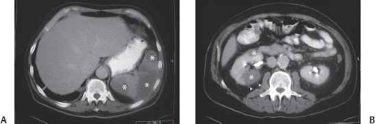
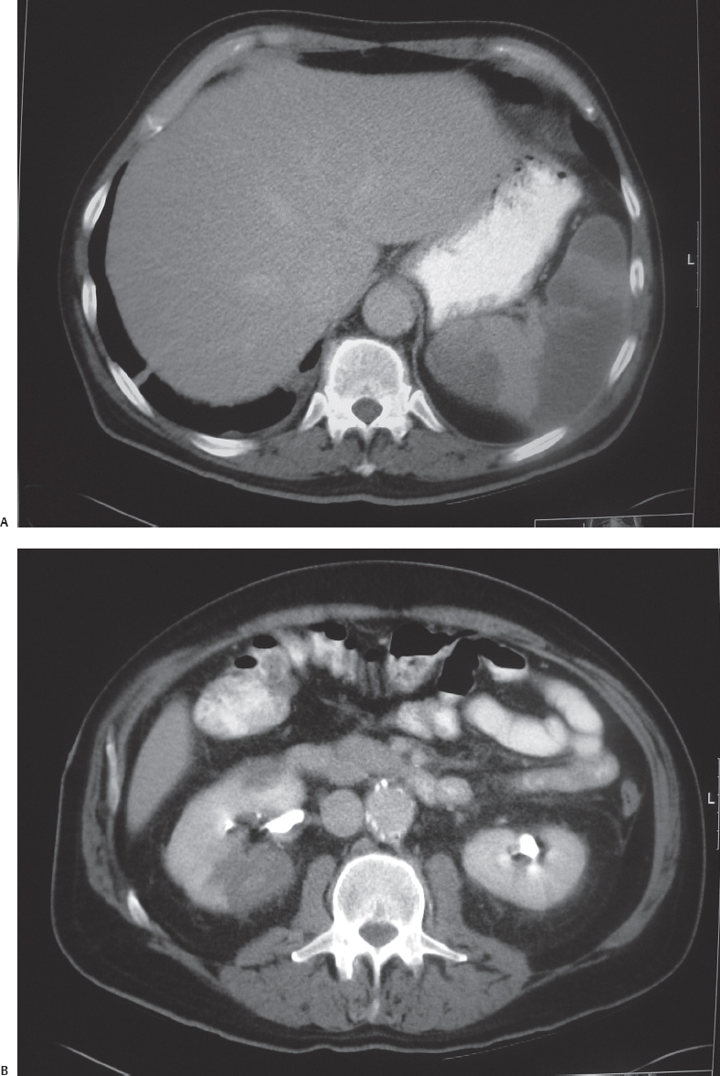
(A) Venous phase contrast-enhanced computed tomography (CT) image at the level of the spleen shows multiple wedge-shaped nonperfused areas (asterisks) in the spleen. (B) Contrast-enhanced CT image at the level of the kidneys in the nephrographic phase shows wedge-shaped perfusion defects (asterisks) in the right kidney. There is a complete absence of perfusion in these areas. Some perinephric fat stranding (arrowheads) is present. Perfusion in the left kidney is normal.
 Differential Diagnosis
• Embolic lobar renal infarcts: Wedge-shaped areas of absent perfusion are characteristic. Multiple infarcts in the kidney and an infarct in the spleen are characteristic of embolic infarction, which in this patient is from a thrombus in the dilated left atrium (not shown) due to atrial fibrillation.
• Acute pyelonephritis: Wedge-shaped perfusion defects are also seen in acute pyelonephritis. There is never a complete absence of perfusion in these areas, and the ischemia is relative. Involvement of other organs is not expected.
• Multiple simple renal cysts:
Stay updated, free articles. Join our Telegram channel

Full access? Get Clinical Tree


