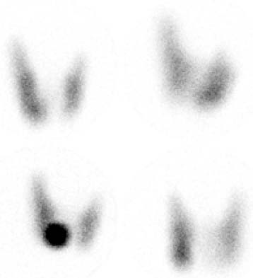
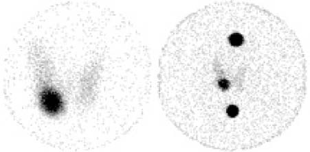
CASE 59 A 35-year-old man presents with an asymptomatic right thyroid nodule noted on routine physical examination. Thyroid function test values are normal. Fig. 59.1 Multiple pinhole collimator views: anterior, right anterior oblique, left anterior oblique, and anterior with “hot” 57Co marker over palpated nodule, 123I. Fig. 59.2 Two pinhole collimator views: anterior without and with anatomic markers (superior marker at chin, inferior marker at sternal notch), 123I after suppression with thyroid hormone. • 0.400 mCi of 123I at 24 hours before scan • Five-minute pinhole collimator views in anterior, right anterior oblique, and left anterior oblique projections, and anterior projection with a “hot” 57Co marker placed over the palpated nodule (Fig. 59.1; look at the images clockwise, beginning with the 57Co marker image in the lower left-hand corner). • After suppression with oral thyroid hormone, repeated anterior pinhole views without and with anatomic markers on the chin (superior marker) and sternal notch (inferior marker) (Fig. 59.2) Figure 59.1 shows a normal-appearing thyroid gland except for minimal asymmetry in tracer uptake. The right lobe appears larger than the left, but this can be a normal variant. The “hot spot” represents the 57Co marker placed on the palpated nodule. Figure 59.2 demonstrates imaging of the same patient after administration of a suppressive dose of thyroid hormone, which “turns off” thyroid-stimulating hormone (TSH) and thus suppresses normally responsive thyroid tissue. The palpated nodule continues to function despite TSH suppression and so is termed autonomous. It is unlikely for a single autonomously functioning nodule to be cancerous, and therefore no further management is required. • Thyroid adenoma • Autonomous adenoma (common) versus thyroid carcinoma (uncommon)
Clinical Presentation
Technique
Image Interpretation
Differential Diagnosis
Stay updated, free articles. Join our Telegram channel

Full access? Get Clinical Tree


