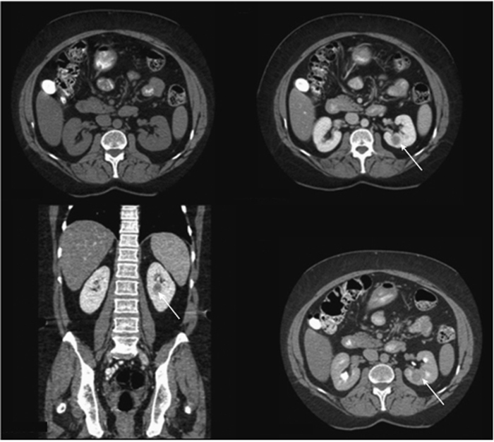
CASE 59 A 56-year-old woman with a history of kidney stones presents with persistent left flank pain. A diagnostic noncontrast computed tomography (CT) scan performed several months earlier showed no evidence of nephrolithiasis and/or renal masses. Fig. 59.1 Single composite image comprising a noncontrast axial image, contrast-enhanced axial and coronal images in portal venous phase, and an axial image in the delayed phase following contrast administration. Noncontrast-enhanced CT demonstrates no discrete focal lesion. In the portal venous phase following contrast enhancement, there is a well-circumscribed mass in the left kidney (arrow), which is hypodense to the surrounding parenchyma. The lesion continues to be visible on the delayed phase (arrow). A single composite image comprises a noncontrast axial image, contrast-enhanced axial and coronal images in the portal venous phase, and an axial image in the delayed phase following contrast administration (Fig. 59.1
Clinical Presentation
Radiologic Findings
![]()
Stay updated, free articles. Join our Telegram channel


Full access? Get Clinical Tree


