6 Management of Complications of Portal Hypertension: Balloon-Occluded Retrograde Transvenous Obliteration, Plug-Assisted Retrograde Transvenous Obliteration, Coil-Assisted Retrograde Transvenous Obliteration, and Balloon-Assisted Antegrade Transvenous Obliteration
Murthy R. Chamarthy and Sanjeeva P. Kalva
6.1 Introduction
Variceal bleeding is the most common complication of portal hypertension. The treatment of varices is dependent on the clinical presentation, the underlying etiology of portal hypertension, and specific characteristics of the varices, such as the size, extent, and location of the varices and the presence of concomitant portosystemic communications. Endoscopic variceal banding is a commonly practiced therapy for symptomatic and asymptomatic esophageal and gastroesophageal varices. Patients in whom endoscopic therapy fails are triaged to portal decompression procedures such as placement of a transjugular intrahepatic portosystemic shunt (TIPS) or other surgical shunts. These therapies are highly effective for esophageal and gastroesophageal varices with low variceal recurrence rates but are less effective in the management of isolated or dominant gastric varices. Gastric varices are treated with endovascular occlusion through a preexisting TIPS, through a new transjugular or transhepatic portal venous approach, or through a systemic venous approach made possible by physiologic portosystemic communications resulting from portal hypertension. The variceal obliterative procedures for gastric variceal occlusion that will be reviewed in this chapter include balloon-occluded retrograde transvenous obliteration (BRTO), plug-assisted retrograde transvenous obliteration (PARTO), coil-assisted retrograde transvenous obliteration (CARTO), and balloon-assisted antegrade transvenous obliteration (BATO).
6.2 Concept of BRTO/PARTO/CARTO
Varices related to portal hypertension are managed through either direct obliteration of the varices or portal decompression. The former approach aims at occluding (through endoscopic therapies) the large submucosal varices, which are prone to bleeding, while sparing the small, nonsubmucosal varices for continued portosystemic shunting. This approach alleviates bleeding and prevents future bleeding from at-risk varices but does little to prevent the development of new varices or alter the progression of portal hypertension. The latter approach aims to decompress the portal vein through the creation of a portosystemic shunt (TIPS or surgical shunt) or the decrease of portal blood flow through splenectomy or splenic embolization. This approach effectively decreases the portal pressure, reduces variceal bleeding, and prevents the development of new varices. However, portosystemic shunt creation may result in hepatic encephalopathy because of portal flow diversion and decreased hepatic detoxification. Systemic venous variceal obliterative procedures aim at direct obliteration of the varices (similar to that achieved with endoscopic therapies) but differ in that they occlude not only the submucosal varices but also the periadventitial large varices and the concomitant portosystemic shunts. These approaches use balloons, vascular plugs, or coils to occlude the systemic venous outflow of the varices and embolize the varices through retrograde injection of liquid embolic materials. In this process, the physiologic shunt is also obliterated. This has profound hemodynamic effects, including decreased rebleeding, improved hepatic encephalopathy and hepatic function, and progression of portal hypertension. Given the excellent outcomes of endoscopic obliteration of esophageal varices, systemic venous variceal obliterative procedures are practiced mainly for gastric varices.
6.3 Gastric Varices
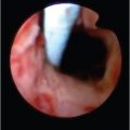
Gastric varices are less commonly encountered than esophageal varices. The prevalence of gastric varices is approximately 17% in cirrhotic patients. 1 Gastric varices may occur along with esophageal varices (gastroesophageal varices) or may be isolated. Isolated gastric varices occur in the presence of splenic or portal vein occlusion. Sarin et al 1 , 2 classified gastric varices into four types based on their endoscopic appearance: GOV1, GOV2, IGV1, and IGV2. GOV1 varices are the most common type (75%) and include esophageal varices extending below the gastroesophageal junction along the lesser curvature of the stomach. GOV2 varices include gastroesophageal varices that extend along the cardia (21%). Isolated gastric varices are rare and may involve the fundus of the stomach (IGV1) or may involve the rest of the stomach excluding the fundus (IGV2).
The risk of bleeding from gastric varices is significantly lower compared to the risk from esophageal varices (10–36% vs. 70–80%); however, the morbidity and mortality associated with gastric variceal bleeding are high. 1 , 2 This is because of the large size and location of gastric varices, which make endoscopic therapies difficult and less effective. Rebleeding rates of gastric varices after endoscopic therapy exceed 70%. In addition, decompression of gastric varices is not effectively achieved with TIPS or surgical shunts because of the low portal pressures associated with gastric varices. The mean portosystemic gradient in the presence of gastric varices is 11.2 compared to 15.5 with esophageal varices. 2 , 3 These low portal pressures result from the presence of concomitant portosystemic shunts that are associated with gastric varices.
6.3.1 Anatomy of Gastric Varices
Venous drainage of the stomach is achieved through the left gastric vein, posterior gastric vein, short gastric veins, and gastroepiploic veins. The left gastric vein drains the distal esophagus, fundus, and the lesser curvature of the stomach, courses along the lesser curvature of the stomach, and drains into the portal vein at the portosplenic confluence. The posterior gastric and short gastric veins drain the body of the stomach and empty into the splenic vein. The gastroepiploic veins drain the body and greater curvature of the stomach and the omentum and drain into the splenic vein from the left gastroepiploic vein or the superior mesenteric vein from the right gastroepiploic vein.
In the presence of portal hypertension, the flow in these afferent veins is retrograde (away from the portal vein) and the afferent veins communicate with various systemic veins via enlarged varices. The gastric veins near the esophagus communicate with the submucosal venous plexus of the lower and midesophagus and drain into the azygos and hemiazygos veins. The veins near the bare area of the stomach communicate with the left inferior phrenic vein. The vertical portion of the left inferior phrenic vein (the ascending phrenic vein) forms the gastrorenal shunt and drains into the left renal vein after joining with the left adrenal vein (most common, 98%) or the left gonadal vein (2%). In addition, the medial horizontal portion of the inferior phrenic vein may drain directly into the inferior vena cava or the right inferior phrenic vein. The lateral horizontal portion of the left inferior phrenic vein receives tributaries from the pericardial vein and intercostal veins. Occasionally, the azygos and hemiazygos veins communicate with the medial horizontal portion of the left inferior phrenic vein.
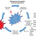
Depending on the flow dynamics, one or more portosystemic communications enlarge and form large shunts. Such shunts include the gastrorenal shunt (gastric varices draining into the left renal vein through the ascending left inferior phrenic vein) and the gastrocaval shunt (gastric varices draining into the medial horizontal left inferior phrenic vein). The former is the most common, occurring in 80 to 85% of patients with gastric varices and in 10 to 15% of patients with portal hypertension. 4 The veins feeding into the varices are often called the “afferents,” and the veins draining into the systemic veins are often called the “efferents”. This nomenclature is extensively used in various classifications as described below and detailed in ▶ Fig. 6.1 and ▶ Fig. 6.2.


6.3.2 Classification of Gastric Varices
Various classifications have been proposed to address the complex anatomy, hemodynamics, and pathophysiology of gastric varices and their relevance to systemic venous obliterative procedures. Irrespective of the classification used for description purposes, it is important to identify the flow dynamics during the procedure and modify the technique of embolization based on the flow pattern, opacification of the varices, and reflux of contrast material into the systemic and/or portal veins.
Kiyosue Classification
In this classification, Kiyosue et al 5 classified the afferents and the efferents into various types to address the complexity of venous flow into and out of the gastric varices. The afferents are classified into three types. In type 1, there is a single afferent vein communicating with the varices. In types 2 and 3, multiple afferent veins exist, and they may directly contribute to the varices (type 2) or may communicate with other afferent veins before contributing to the varices (type 3). Depending on the flow pattern and hemodynamic changes with ongoing embolization, there is a risk of nontarget spread of embolic materials into the portal vein with types 2 and 3 afferent systems. An antegrade balloon occlusion or embolization of the afferent veins may allow successful treatment of the varices in these scenarios.
The efferent system is classified into four types depending on the number and caliber of the efferent veins. In type A, there is a single efferent draining vein, commonly a gastrorenal shunt. In type B, multiple efferent draining veins exist, but there is a single large shunt with multiple small efferents that do not pose any significant risk of nontarget systemic embolization during the procedure. Type B is subclassified into B1 (small low flow), B2 (medium-sized low flow), and B3 (large high flow without discrete shunt). In type C, multiple large shunts exist, with a high risk of nontarget systemic embolization unless the shunts are actively occluded before the varices are treated (C1, smaller second shunt; C2, larger sizable second shunt). In type D, there is no sizable systemic venous communication to allow systemic venous variceal obliteration. A recent modification of type D, known as Saad–Kiyosue Type D2, takes into consideration the uncommon portosystemic shunts (through efferents such as the azygos, hemiazygos, pericardial, or intercostal veins) that allow for systemic venous variceal obliterative procedures.
Hirota Classification
In the Hirota classification system, the gastric varices are classified into different types based on the degree of visualization during balloon-occluded retrograde venography (BORV). 6 In grade 1, the gastric varices are well opacified without visualization of any collateral veins. In grade 2, there are a few small collaterals but the gastric varices are adequately opacified for > 3 minutes. In grade 3, the gastric varices demonstrate medium to large collaterals with only partial opacification and quick disappearance of contrast material (< 3 min). In grade 4, there are large collateral veins without opacification of the gastric varices. Grade 5 indicates a shunt that is not amenable to balloon occlusion; this is the most common cause of technical failure.
Fukuda–Hirota Hemodynamic Classification
In the Fukuda–Hirota hemodynamic classification system, the flow pattern in the gastric and esophageal varices and concomitant portosystemic shunt opacification during arterioportography are taken into consideration. 7 In type 1, there is a predominant left-sided portosystemic shunt resulting in fundal gastric varices without esophageal varices. In type 2, the esophageal varices are opacified but are not connected to and are independent of gastric variceal drainage. Type 3 is represented by complex gastroesophageal varices with a gastrorenal shunt as the primary outflow. Type 4 involves gastric varices supplied by the left gastric vein.
Matsumoto Hemodynamic Classification
Based on left gastric arteriography, Matsumoto et al 8 classified gastric varices into type I and type II based on the presence or absence of communication between the left gastric artery and the gastrorenal shunt, respectively. The varices are subdivided further into a and b groups based on the hepatopetal or hepatofugal blood flow within the left gastric vein. The type Ib group (presence of communication from the left gastric artery to the gastrorenal shunt and presence of hepatofugal flow within the left gastric vein) is associated with the highest risk of worsening esophageal varices subsequent to BRTO.
Saad–Caldwell Classification
The Saad–Caldwell classification is based on the type of varices (gastroesophageal vs. gastric) and the presence of a gastrorenal shunt (type a, without gastrorenal shunt; type b, with gastrorenal shunt). 9 Type I gastric varices correlate with Sarin′s GOV1 class, and the management of these varices is similar to that used for esophageal varices. TIPS placement is preferred for type Ia, whereas a TIPS procedure with left gastric vein embolization is suggested for type Ib. Type II gastric varices correlate with Sarin′s IGV1 class. TIPS placement with afferent venous embolization is suggested for type IIa; BRTO is suggested for type IIb, as TIPS placement would be less effective in the presence of a high-flow gastrorenal shunt. Type III gastric varices are complex cardiofundic gastric varices associated with esophageal varices. TIPS placement and antegrade variceal embolization are suggested for type IIIa, and BRTO with or without TIPS placement is suggested for type IIIb. Type IV gastric varices occur in the presence of splenic or portal vein thrombosis. BRTO can be considered if the portal vein is patent and a gastrorenal shunt is present. Otherwise, splenic artery embolization is recommended.
6.4 BRTO
6.4.1 Indications and Contraindications
The indications and contraindications for BRTO procedures in the management of gastric varices are summarized in ▶ Table 6.1 and ▶ Table 6.2. 10 The main indication for BRTO/PARTO/CARTO is the presence of large gastric or gastroesophageal varices that are at risk of bleeding or have already bled and are recalcitrant to other therapies. The presence of refractory hepatic encephalopathy secondary to a large gastrorenal or gastrocaval shunt is another indication for this procedure. Gastroesophageal varices with predominant afferent supply from the left gastric vein might benefit from a TIPS placement, which can be performed in conjunction with a BRTO/BATO procedure, especially in the setting of predominant esophageal varices, refractory hydrothorax, and ascites. 11 Splenic artery embolization is performed in combination with BRTO or alone (for type D varices) to decrease the portal pressure in cases with splenic vein thrombosis and splenomegaly.
The BRTO procedure is contraindicated when the main portal vein is occluded and the gastrorenal shunt is the sole pathway for physiologic portal decompression. Occlusion of the gastrorenal shunt in this setting would result in portal flow stagnation and mesenteric venous thrombosis. In these circumstances, the varices may be obliterated if an additional portal decompression surgery is performed. The ability of cavernous transformation of the portal vein to handle the increased portal circulation from a BRTO procedure is unknown and debatable. Partial thrombosis of the portal vein may in fact benefit from the procedure, with increased flow and decreased stasis. Splenic vein thrombosis is a partial contraindication; BRTO can be performed in conjunction with splenic artery embolization to address the problem of portal hypertension. Coexisting esophageal varices or refractory hydrothorax or ascites are relative contraindications; in such cases, combination therapy with TIPS placement might be considered. A conventional BRTO procedure cannot be performed in the absence of a gastrorenal shunt. In this setting, BATO (type D1) or a nonconventional venous access (type D2) is needed for obliteration. The procedure is also contraindicated when there is a high risk of nontarget embolization of systemic or portal veins.
Related posts:


Stay updated, free articles. Join our Telegram channel

Full access? Get Clinical Tree


