9 Interventional Management of Benign Refractory Ascites
Louis G. Martin
9.1 Introduction
Those of us who saw the movie Amadeus will never forget the opening scene of the carriage traveling through a cold, late-night thunderstorm, bringing the priest to the deathbed of Antonio Salieri, who confesses his role in the death of Wolfgang Amadeus Mozart. Mozart actually died at home on a calm, mild day in Vienna after a short illness—and without Salieri′s intervention. A much more dramatic demise actually belonged to Ludwig van Beethoven, who died in a hepatic coma during a violent thunderstorm with snow flurries and hail. According to the case notes of Anton Wawruch, the physician caring for Beethoven at the time of his death in 1827, “Beethoven opened his eyes for the first time in 2 days, raised his right hand in a clenched fist, and stared up into the heavens with a grim and threatening look on his face, and then expired.” 1 , 2 Beethoven is undoubtedly the most celebrated patient to have suffered from refractory ascites (RA). He received paracentesis at least four times during the 2 months that preceded his death (reportedly up to 20 L per session). Beethoven′s paracentesis was performed without anesthesia by the introduction of a glass tube through a surgical incision, as was typical practice at the time. 3 , 4
Abdominal paracentesis is one of the oldest surgical procedures recorded. It was described in a single passage of the Hippocratic Writings, in which the author says “that a puncture of the abdomen near the umbilicus or in the region of the flank should be made either by knife or cautery and that only a small amount of fluid should be allowed to flow at a time.” 5 This is a very sophisticated passage despite its brevity; the methodology described lessens the likelihood of trauma to the recanalized umbilical vein, other collateral veins on the surface of the abdomen, and the branches of the superficial epigastric artery and vein. Additionally, this technique reduces the possibility of causing vasomotor instability. In his extant work De Medicina, Aulus Cornelius Celsus (c. 25 BC to c. 50 AD) described in detail the technique of paracentesis, recommending the use of a bronze tube with a flange collar to prevent the tube from slipping into the patient′s peritoneal cavity. He noted that Erasistratus of Alexandria opposed paracentesis because of its hazards. Six centuries later, Paul of Aegina warned that sudden evacuations have “immediately killed the patient.” 6
Large-volume paracentesis (LVP) was the only treatment available for ascites for more than 2,000 years. Salt restriction was not attempted or even suggested until the early 1900s. Although mercury was advocated as a diuretic in the sixteenth century, there is no record of mercury being used to treat ascites until organic mercurial agents were introduced after World War II. The discovery of sulfanilamide-induced sodium bicarbonate diuresis in the late 1940s ushered in a new age of clinically effective diuretics, which began in the 1950s with the introduction of chlorothiazide and spironolactone, the first orally effective agents to mobilize sodium chloride. 3 , 7
In this chapter, we will review the pathogenesis of cirrhotic RA and the differences between compensated and uncompensated cirrhosis. We will discuss the many treatment options available for RA and summarize their benefits, contraindications, and complications. Finally, we will propose a treatment algorithm that meets the needs of the patient and satisfies the goals of the interventional radiologist, hepatologist, and transplant surgeon. References to malignant ascites will be included only when they serve to clarify issues pertaining to cirrhotic ascites.
9.2 Pathogenesis of Cirrhosis
The elements active in the pathogenesis of cirrhosis are not completely known. Its present state of understanding has progressed through: the backward flow theory, the forward flow theory, and the vasodilatation-hyperdynamic circulation theory. Each of these theories has added information to previous theories but has not supplanted them. The backward flow theory postulates that portal hypertension is due solely to increased resistance caused by hepatic sinusoidal fibrosis. This is observed in early cirrhosis before portal flow is diverted into the collateral circulation. The forward flow theory acknowledges the role of increased portal resistance but recognizes the equal importance of increased splanchnic blood flow, providing a rationale for the use of vasoconstrictors in this patient population. The vasodilatation-hyperdynamic circulation theory postulates that the “trigger” of the hyperdynamic syndrome is in the action of multiple vasodilators, primarily nitric oxide and endothelial growth factor, in the splanchnic bed and is activated by the initial increase in portal pressure related to sinusoidal hypertension 8 , 9 , 10 (▶ Fig. 9.1).

9.3 Compensated versus Decompensated Cirrhosis
Normally, there is only 50 to 150 mL of fluid in the peritoneum; increased fluid as a result of trauma, infection, neoplasia, or portal hypertension is initially drained via the lymphatics. When this drainage is insufficient, dietary and medicinal assistance may be required. Biliary cirrhosis is usually an indolent process, often taking 8 to 10 years to progress from a compensated to decompensated state. Early clinical signs and manifestations of compensated cirrhosis are pruritus, persistent fatigue, and pain in the right upper quadrant of the abdomen. 11 The onset of decompensation is defined as the presence of one or more of the following complications: jaundice, ascites, spontaneous bacterial peritonitis (SBP), hepatic encephalopathy (HE), hepatorenal syndrome (HRS), variceal hemorrhage, and cancer. 12
Cirrhosis is the most common cause of ascites, accounting for nearly 85% of all cases. Similarly, ascites is the most frequent decompensating event, occurring in 60% of cirrhotic patients within 10 years of disease onset. The presence of ascites is associated with 50% mortality within 3 years and 50% mortality within 1 year once the ascites has become refractory to diuretics. 13 An increase in hepatic sinusoidal fibrosis, increase in portal vein pressure, and formation of a hyperdynamic splanchnic circulation are important factors involved in the formation of ascites. Rupture of either the liver capsule and/or its surface lymphatics allows for high protein fluid leak into the peritoneal space, which is the crucial event in the formation of ascites. In the setting of progressive vasodilatation, the intravascular volume and cardiac output increase to maintain arterial perfusion pressure. With progression of the disease, vasodilatation is accentuated, and cardiac output continues to increase. Eventually, the cardiac response is not enough to maintain perfusion pressure, the renal blood flow drops, and renal failure develops. 8 The additional increase in levels of endogenous vasoactive compounds such as renin, angiotensin, aldosterone, norepinephrine, vasopressin, and antidiuretic hormone results in increased ascites accumulation and sodium retention. 14 , 15
The most important markers of decompensated cirrhosis are bleeding and ascites. Additional complications of cirrhosis can include edema, SBP, HE, HRS, hepatopulmonary syndrome, hypersplenism, and liver cancer (hepatocellular carcinoma [HCC], which is the third leading cause of cancer mortality worldwide). The 5-year cumulative risk for the development of HCC in patients with cirrhosis ranges from 5 to 30%. 16 , 17 , 18 , 19
Survival of cirrhosis depends on the degrees of portal hypertension, liver insufficiency, and circulatory dysfunction. Prognostic factors for survival include the Model for End-Stage Liver Disease (MELD) score, 20 portosystemic pressure gradient ≥ 10 mm Hg, 21 and increased body mass index. 22
Successful treatment of ascites is dependent on accurate diagnosis of its cause. Because sodium and water retention is the basic abnormality leading to ascites formation, restricting sodium intake and enhancing sodium excretion are the mainstays of ascites treatment. Patients with cirrhosis and ascites must limit sodium intake to 2 g/d. Enhancement of sodium excretion can be accomplished with oral diuretics. The recommended initial dose is spironolactone 100 to 200 mg/d and furosemide 20 to 40 mg/d; the dosages of these medications can be increased if the response is not adequate to control ascites. The usual maximum doses are spironolactone 400 mg/d and furosemide 160 mg/d. The recommended weight loss in patients without peripheral edema is 300 to 500 g/d. There is no limit to the daily weight loss of patients with edema. Approximately 90% of patients respond well to medical therapy for ascites.
9.4 Refractory Ascites
Approximately 5 to 10% of patients with cirrhosis develop RA. Once the diagnosis of RA is established, the expected 1-year survival is approximately 50%. Predictors of poorer survival in patients with RA include low protein levels in the ascitic fluid, elevated Child–Pugh score, previous SBP, heavy alcohol consumption (> 80 g/d in men and > 40 g/d in women), older age, the presence of HCC, and the presence of diabetes. 13 , 23 , 24 , 25 , 26
The diagnostic criteria for RA as established by the International Ascites Club 27 and accepted by the American Association for the Study of Liver Disease 28 are as follows: RA is defined as fluid overload that is unresponsive to a sodium-restricted diet (≤ 50 mEq/d) and diuretic treatment or that recurs rapidly after therapeutic paracentesis. Diuretic-resistant RA is defined as ascites accumulation that is not controlled by a sodium-restricted diet plus maximum doses of spironolactone and furosemide (other alternatives include bumetanide 4 mg/d or an equivalent dose of other loop diuretics for at least 1 week). Diuretic-intractable RA is defined as RA that cannot be treated with the maximal dose of diuretics because of diuretic-induced complications.
Lack of response in patients with RA is defined as weight loss less than 200 g/d during the last 4 days of intensive diuretic treatment and urinary sodium excretion less than 50 mEq/d. Early ascites recurrence is defined as the reappearance of moderate (grade 2) to massive or tense (grade 3) ascites within 4 weeks of initial mobilization. Reaccumulation of ascites within 2 to 3 days of paracentesis should not be considered early ascites because it represents a shift of interstitial fluid to the intraperitoneal space. 27
Diuretic-induced complications are a frequent occurrence even in hospitalized patients. These complications include the development of HE in the absence of other precipitating factors (25% of patients), increase of serum creatinine to more than 2 mg/dL and/or by more than double the base value (20% of patients), decrease in serum sodium concentration by more than 10 mEq/L to a level lower than 125 mEq/L (30% of patients), and diuretic-induced hypokalemia (decrease of serum potassium to < 3 mEq/L) or hyperkalemia (increase of serum potassium to > 6 mEq/L) despite the use of appropriate measures to correct serum potassium levels. 27 , 29
RA is not only a problem in patients with decompensated cirrhosis; one single-center retrospective study found that RA occurred in 62 (5.6%) of 1,058 patients who underwent orthotopic liver transplant (OLT). 30 Successful treatment of RA in the posttransplant patient resulted in significantly improved survival (p = 0.00001). Therefore, the interventionalist must search for and correct treatable causes of RA in this patient cohort. Anastomotic stenosis of the inferior vena cava, hepatic veins, and portal veins may also occur in 3 to 7% of patients after OLT. Technical success rates of 94 to 100% and a clinical success rate of 73% have been reported after percutaneous treatment of post-OLT anastomotic strictures. 31 , 32 In the absence of anastomotic stenosis, transjugular intrahepatic portosystemic shunt (TIPS) placement 33 and splenic artery embolization 34 have been shown to have benefit in the treatment of RA and as a bridge to retransplantation.
9.5 Conditions Coexisting with or Complicating Refractory Ascites
9.5.1 Spontaneous Bacterial Peritonitis
SBP is the most frequent infection to occur in patients with RA affecting 3.5% of outpatients and 12% of hospitalized patients. As many as 65% of patients with borderline renal function, ascitic fluid protein level ≤ 1.5 g/dL, Child–Pugh score ≥ 9, and bilirubin level ≥ 3 mg/dL may experience SBP within 1 year of interventional treatment. 35 The 1-year probability of SBP recurrence is 70%, with a corresponding survival rate of 50 to 80%. 23 , 36 , 37 SBP is caused by gut bacteria or bacterial products crossing from the intestinal lumen into the blood or ascitic fluid. This bacterial translocation occurs as a result of overgrowth of intestinal bacteria in an immunologically impaired patient with cirrhosis and with increased intestinal permeability. 38
A routine cell count of fluid obtained from diagnostic or therapeutic paracentesis should always be performed to rule out SBP. The presence of an ascitic fluid polymorphonuclear leukocyte count ≥ 250 cells/mm3 without evidence of a surgically correctable source of infection confirms the diagnosis of SBP. A dipstick specifically designed for ascitic fluid and calibrated to 250 cells/mm3 is reported to have a 95% sensitivity and 79.6% specificity for SBP detection; however, this test is not effective in bloody, chylous, or bilious fluid. 39 Delaying treatment until bacteria grow in ascitic fluid culture may result in the death of the patient. Empiric intravenous administration of a third-generation cephalosporin antibiotic (2 g twice daily) should be initiated immediately, and the antibiotic should be replaced when necessary (depending on the results of ascitic fluid culture and sensitivity tests). Additionally, the high risk of SBP reinfection can be significantly reduced by prophylactic oral administration of norfloxacin 400 mg/d (p = 0.0063). 40
9.5.2 Hepatorenal Syndrome
HRS is defined as the occurrence of renal failure in a patient with advanced liver disease in the absence of an identifiable cause of renal failure; thus, the diagnosis is one of exclusion. 29 However, recent evidence suggests that HRS is preceded and intimately related to cardiac dysfunction. A low cardiac index (CI) has been identified as an independent predictor of the development of HRS. 41 In a study of patients with cirrhosis and ascites but without HRS, patients with a CI lower than 1.5 L/min/m2 demonstrated significant reductions in stroke volume, stroke volume index, heart rate, systemic vascular resistance, renal blood flow, glomerular filtration rate, and serum aldosterone and an increase in serum creatinine versus patients with a CI higher than 1.5 L/min/m2. Mortality at 3, 6, and 9 months was also significantly higher in patients with a lower CI, a result not predicted by differences in MELD scores. 42
The inability to excrete sodium is not apparent until ascitic fluid accumulates in the peritoneal cavity. The renal circulatory bed attempts to compensate for the hyperdynamic cirrhotic state through reactive vasodilatation, which results in retention of sodium and water. As decompensation progresses, vasodilatation is accentuated, and the cardiac output continues to increase. When the cardiac response is unable to maintain perfusion pressure, renal vasoconstriction occurs and renal failure develops. 8 , 41 Ginès and Schrier 43 concluded that HRS is probably the final consequence of extreme underfilling of the arterial circulation secondary to arterial vasodilatation in the splanchnic vascular bed. As a result, arterial pressure must be maintained by activation of vasoconstrictor systems (i.e., the renin–angiotensin–aldosterone system, sympathetic nervous system, and, in late stages, nonosmotic antidiuretic hormone). Thus, in patients with HRS, the renal circulation and most extrasplanchnic vascular beds are vasoconstricted.
There are two types of HRS. Type 1 is characterized by rapidly progressive deterioration of renal function, resulting in a 24-hour creatinine clearance of less than 20 mL/min and a serum creatinine level greater than 2.5 mg/dL in less than 2 weeks. Bacterial infections, gastrointestinal hemorrhage, major surgical procedures, and acute-on-chronic liver failure are the most frequent precipitating events. In patients with type 1 HRS, hospital survival is less than 10%, and the expected median survival time is only 2 weeks. 44 The European Association for the Study of the Liver recommends that terlipressin (1 mg/4–6 h intravenous bolus) plus albumin should be used as first-line therapy for type 1 HRS. This treatment regimen aims to improve renal function sufficiently to decrease the serum creatinine level to less than 1.5 mg/dL. The terlipressin dose should be increased in a stepwise manner to a maximum of 2 mg/4 h if serum creatinine does not decrease by at least 25% in 3 days. Terlipressin is not available in the United States; potential alternatives include norepinephrine or midodrine plus octreotide in combination with albumin. 45 Vasoconstrictors, mainly terlipressin and beta-blockers, have resulted in improved renal function in some patients with HRS type 1; however, these agents should be used with caution as many patients in this population have a reduced CI and so a further decrease in systemic blood flow and oxygen transport may be deleterious. 42
Type 2 HRS progresses more slowly, with a serum creatinine level ranging from 1.2 to 2.5 mg/dL; median survival in these patients is 6 months. Type 2 HRS is mainly characterized by the presence of RA. Albumin infusion has been shown to prevent type 2 HRS and improve survival, especially in the setting of SBP. 46 Pentoxifylline, a phosphodiesterase inhibitor, has been shown in a randomized trial to be superior to placebo in preventing type 2 HRS in patients with cirrhosis, ascites, and creatinine clearance levels between 41 and 80 mL/min. 47
During the progression of liver disease, there is concomitant progression of renal perfusion disturbances and renal sympathetic overactivity. 48 Recently, there has been renewed interest in sympathetic denervation as a treatment for the renal vasoconstrictive complications seen in HRS. Selective catheterization and tracer kinetic techniques have demonstrated that the increased circulating norepinephrine in patients with cirrhosis is due to enhanced sympathetic nervous system activity in the kidneys and other organs. 49 Surgical lumbar sympathectomy has led to improvements in renal function in several patients with HRS. 50 Placement of TIPS has also been reported to improve renal function in patients with type 1 HRS; although these data are encouraging, they are inconclusive, and many patients have contraindications to TIPS placement. 51 , 52 These treatment options will be discussed in more detail later in this chapter.
9.5.3 Hepatic Encephalopathy
HE describes a spectrum of potentially reversible neuropsychiatric abnormalities seen in patients with liver dysfunction and/or portosystemic shunting. Overt HE develops in 30 to 45% of patients with cirrhosis and in 10 to 50% of patients with TIPS placement (▶ Table 9.1). 53 , 54 The International Society for Hepatic Encephalopathy and Nitrogen Metabolism consensus document defines the onset of disorientation or asterixis as the onset of overt HE. 55 Some patients with HE have subtle findings that may only be detected using specialized tests; this is known as minimal HE and occurs in up to 80% of patients with cirrhosis. 56 Refractory HE is defined as encephalopathy that recurs or persists despite the use of appropriate medical treatment. The prognosis for patients with overt HE is poor; a population-based cohort study reported a 1-year survival rate of 36%. 57 OLT is the ultimate treatment for refractory HE; however, the shortage of available donor organs markedly limits this treatment option. Alternative therapies such as shunt occlusion or reduction can be used to control symptoms and serve as a bridge therapy to OLT. 58
The pathogenesis of HE is poorly understood. The most widely accepted theory is that the astrocyte, the only cerebral cell capable of metabolizing ammonia, is morphologically altered by hyperammonemia. Ammonia detoxification in astrocytes leads to accumulation of glutamine, which is the main cause of astrocyte swelling. Ammonia is converted to carbamoyl phosphate, which then enters the urea cycle to be either incorporated into amino acids or excreted in the urine. Increased production and decreased excretion of ammonia are associated with portal hypertension. In patients with normal hepatocyte function, 80 to 90% of ammonia is excreted through first-pass metabolism. Although ammonia is released from several tissues, including kidney and muscle tissue, its highest concentration is in the portal vein, where it is derived from colonic bacteria and metabolism of glutamine in the small bowel. There is evidence that elevated intracellular ammonia results in altered neurotransmission by agonizing gamma-aminobutyric acid (GABA) and by causing cerebral energy failure. Among patients with HE, 90% have elevated serum ammonia concentrations; however, there is poor correlation between the venous concentration of ammonia and the grade of HE, lending credence to other theories regarding HE pathogenesis. 59 , 60 Discussion of these theories is beyond the scope of this review; the reader is encouraged to investigate the relevant references. 61 , 62 , 63 , 64 , 65
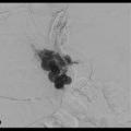
Up to 80% of patients with cirrhosis have subtle findings of HE that may only be detected using specialized tests. These are considered to have mild HE, which may affect 30 to 70% of patients with cirrhosis. 56 , 57 Symptomatically overt HE develops in 30 to 45% of patients with cirrhosis and in 10 to 50% of patients treated by placement of a TIPS shunt. 53 , 54 The International Society for Hepatic Encephalopathy and Nitrogen Metabolism defines the onset of disorientation or asterixis as signaling the onset of overt HE. Refractory HE is defined as a recurrent or persistent encephalopathy despite appropriate medical treatment. HE is reversible; its treatment relies on suppressing the production of the toxic substances in the intestine, which is most commonly achieved with the laxative lactulose or with nonabsorbable antibiotics. Treatment of any reversible underlying conditions such as bleeding, infection, renal failure, constipation, use of psychotropic drugs, and electrolyte abnormalities may improve the symptoms. 59 Surgical approaches to reduce the intestinal production of ammonia, such as colectomy or colon exclusion procedures, were used in the past for patients with HE refractory to other measures. Although these approaches were successful, the operative and postoperative morbidity and mortality rates were high; today, OLT should be considered for these patients. 66 , 67 There is early evidence that partial splenic artery embolization (PSAE) can have a positive effect on the control of HE. In one study, 25 patients with HE were divided into 2 groups: 14 patients underwent transportal obliteration and/or balloon-occluded retrograde transvenous obliteration of porto-systemic shunts followed by PSAE, and 11 patients underwent only transportal obliteration and/or balloon-occluded retrograde transvenous obliteration of portosystemic shunts without PSAE. Serum ammonia levels and HE grades were lower in patients treated with PSAE than in patients not treated with PSAE at 6, 9, 12, and 24 months after treatment. 68
Related posts:
 6 Management of Complications of Portal Hypertension: Balloon-Occluded Retrograde Transvenous Obliteration, Plug-Assisted Retrograde Transvenous Obliteration, Coil-Assisted Retrograde Transvenous Obliteration, and Balloon-Assisted Antegrade Transvenous Obliteration
 15 Hepatocellular Carcinoma: Staging and Clinical Management
 12 Malignant Obstructive Jaundice
 23 Yttrium-90 Radioembolization of Malignant Liver Tumors
 27 Image-Guided Colorectal Obstruction Management
 28 Obesity and Bariatric Embolization
Stay updated, free articles. Join our Telegram channel

Full access? Get Clinical Tree


