Case 60
 Clinical Presentation
A 32-year-old woman with a palpable pelvic mass.
 Imaging Findings
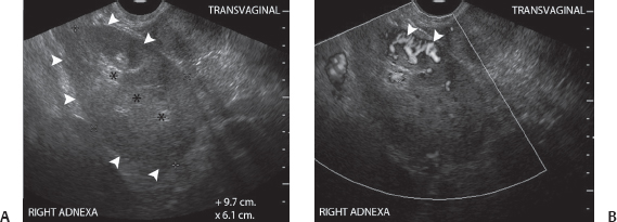
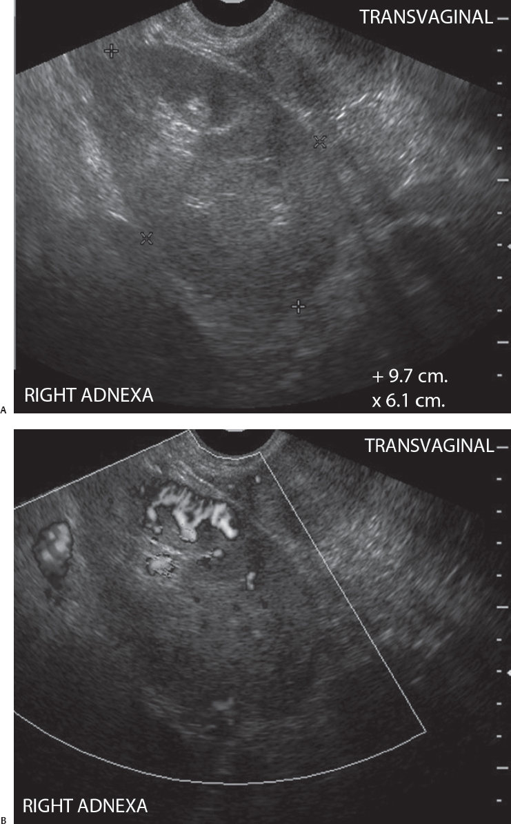
(A) Transvaginal ultrasound image of the pelvis shows a 9.7 × 6.1-cm mass with a central hyperechoic area (black asterisks) surrounded by a hypoechoic rim (arrowheads). (B) Transvaginal ultrasound image of the pelvis with power flow shows a well-organized radial vascular pattern (arrowheads) in parts of the hypoechoic portion of the mass shown in Figure A.
 Differential Diagnosis
• Pelvic kidney: A “mass” with central echoes surrounded by a thick hypoechoic mantle is the characteristic appearance of a pelvic kidney. The well-organized radial vessels in the hypoechoic mantle also strongly suggest that the “mass” is a pelvic kidney rather than an adnexal neoplasm.
• Ovarian dermoid cyst: A sebaceous plug composed of fat and sebum in an ovarian dermoid also appears as an echogenic area within a hypoechoic mass in the adnexa. The mass does not show the layered architecture of the kidney. Acoustic shadowing is expected distal to the sebaceous plug. No organized vessels are seen in the hypoechoic area of the cyst.
Stay updated, free articles. Join our Telegram channel

Full access? Get Clinical Tree


