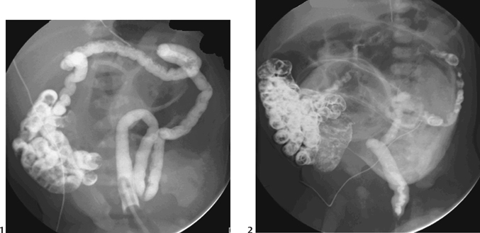
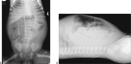
60 Microcolon/Meconium Ileus

CASE 60


Clinical Presentation


Term neonate presents with a distended abdomen and failure to pass meconium after 48 hours.


image

Figure 60A


Radiologic Findings


Non-ionic contrast enema (Fig. 60A1) in a 2-day-old girl demonstrates a microcolon. Reflux of contrast has occurred into mildly dilated loops of distal ileum, containing meconium plugs, but does not reach the more proximal dilated air-filled loops. Further reflux was achieved during a repeat examination the following day (Fig. 60A2), with opacification of significantly more dilated loops in the mid-ileum, containing further inspissated meconium.


Diagnosis


Meconium ileus


Differential Diagnosis


  • Low gastrointestinal (GI) obstruction on abdominal radiograph-wide differential

    • Ileal atresia
    • Hirschsprung’s disease
    • Functional immaturity/left colon syndrome
    • Anorectal anomaly
    • Colonic atresia
    • Sepsis/electrolyte disturbance
    • Megacystis-microcolon-hypoperistalsis syndrome (rare)

  • Microcolon on enema

    • Ileal atresia—microcolon with reflux of contrast into nondilated distal ileum. If contrast reaches the atretic segment, an abrupt convex termination is seen, with residual unopacified dilated proximal small bowel loops.
    • Megacystis-microcolon-hypoperistalsis syndrome (rare)—transient neonatal microcolon in association with a large-volume bladder and a variable degree of upper renal tract dilatation. Malrotation is common.
    • Total colonic Hirschsprung’s disease (may present as a small-caliber colon, although not usually as small as a “microcolon”)

Discussion


Background


Meconium ileus is a neonatal obstruction of the ileum by abnormally viscid, inspissated pellets of meconium.


Etiology


  • Almost all cases are associated with an underlying diagnosis of cystic fibrosis (CF). Conversely, meconium ileus is the initial presentation of 10 to 15% of CF cases.
  • Rare cases have been described in association with partial pancreatic aplasia or stenosis of the pancreatic ducts.

Clinical Findings


Low GI obstruction—abdominal distension, failure to pass meconium, and sometimes vomiting — within the first 48 hours of life


Associated Conditions


Complications


image

Figure 60B Supine (1) and cross-table lateral (2) abdominal x-ray of a 2-day-old girl showing generalized bowel dilatation with a mottled appearance of air mixed with meconium, in this case more prominent in the left flank. Enema confirmed meconium ileus with an underlying diagnosis of CF.


Pathology

Stay updated, free articles. Join our Telegram channel

Dec 21, 2015 | Posted by in PEDIATRIC IMAGING | Comments Off on 60 Microcolon/Meconium Ileus

Full access? Get Clinical Tree

Get Clinical Tree app for offline access