Case 61
 Clinical Presentation
A 36-year-old woman with a palpable lower abdominal mass.
 Imaging Findings
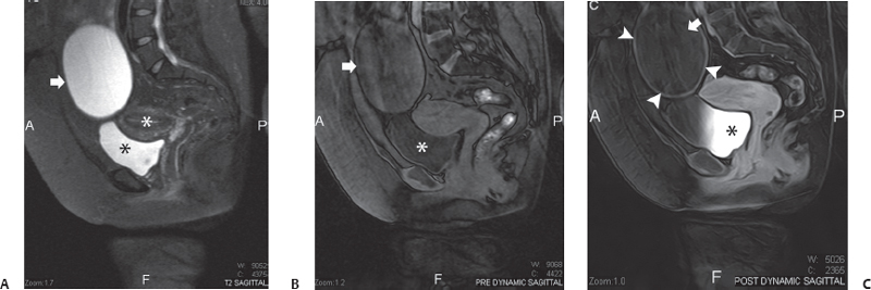
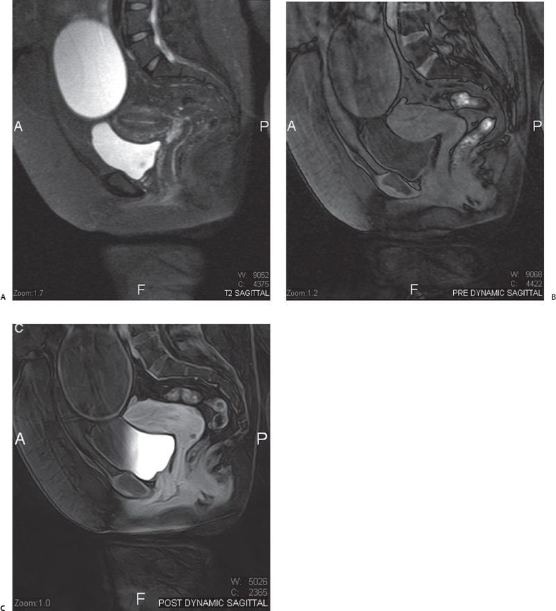
(A) Sagittal midline fat-saturated T2-weighted image of the pelvis shows a large mass of fluid signal intensity (arrow) above a normal urinary bladder (black asterisk) and normal uterus (white asterisk). There are no septa, and the wall is thin. No soft-tissue projections are seen arising from the wall. (B) Sagittal midline fat-saturated precontrast T1-weighted image of the pelvis and lower abdomen at the same level as in Figure A shows the same mass (arrow). The contents have a low signal similar to that of the urine in the urinary bladder (asterisk). (C) Sagittal midline fat-saturated postcontrast T1-weighted image of the pelvis and lower abdomen at the same level as in Figure B shows that the cystic mass (arrow) has a regular, mildly enhancing wall (arrowheads). No enhancing soft-tissue masses or septa are present. Excreted contrast is seen in the urinary balder (asterisk).
 Differential Diagnosis
• Ovarian cyst with no imaging signs of malignancy:
Stay updated, free articles. Join our Telegram channel

Full access? Get Clinical Tree


