CASE 61
Clinical Presentation
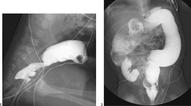
Contrast enema was performed on a 2-day-old term baby who had failed to pass meconium.
Figure 61A
Radiologic Findings
An abrupt transition zone is seen at the rectosigmoid junction (Fig. 61A1), with mild dilatation of the remainder of the colon (Fig. 61A2).
Diagnosis
Hirschsprung’s disease
Differential Diagnosis
- Neonatal low obstruction:
- Functional immaturity/left colon syndrome
- Meconium ileus
- Ileal atresia
- Colonic atresia
- Megacystis-microcolon-intestinal hypoperistalsis syndrome (rare)
- Constipation in the older child:
- Functional megarectum
- Motility disorders (primary and secondary visceral myopathies and neuropathies)
- Metabolic disorders (e.g., hypothyroidism)
- Histologic findings:
- Rare—Chagas’ disease (destruction of ganglion cells)
Discussion
Background
Hirschsprung’s disease, named eponymously following a report of two cases of megacolon in 1887, is due to aganglionosis of the distal bowel. It occurs with an incidence of ~1:4500 live births and demonstrates an overall 4:1 male-to-female ratio.
Etiology
- The primary abnormality is failure of the normal craniocaudal migration of vagal neural crest cells between weeks 5 and 12 of gestation. This results in distal absence of ganglion cells in the myenteric (Auerbach) plexus and submucosal (Meissner) plexus of the bowel wall. The agan-glionic segment begins at the anal sphincter and extends proximally for a varying length of the colon. It is continuous, the presence of “skip” lesions being extremely rare.
- The physiology of the disease is more complex than pure aganglionosis. Abnormalities of both adrenergic and cholinergic fibers can be demonstrated, and function of the internal anal sphincter is also abnormal.
Classification
- Short segment disease (70% cases): The aganglionic segment involves the rectum and distal sigmoid colon.
- Long segment disease (25%): The aganglionic segment extends to the splenic flexure or transverse colon.
- Total colonic aganglionosis (Zuelzer-Wilson syndrome) (5%): The entire colon is involved with occasional extension into the small bowel. Total colonic disease demonstrates a strong familial tendency.
- Ultrashort segment disease: This remains a controversial entity in which the abnormality is limited to the anal sphincter. A manometric and clinical diagnosis, there are no abnormal radiologic or histologic findings.
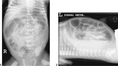
Figure 61B Supine (1) and cross-table lateral (2) abdominal x-ray demonstrating multiple dilated loops of bowel with air-fluid levels.
Clinical Findings


