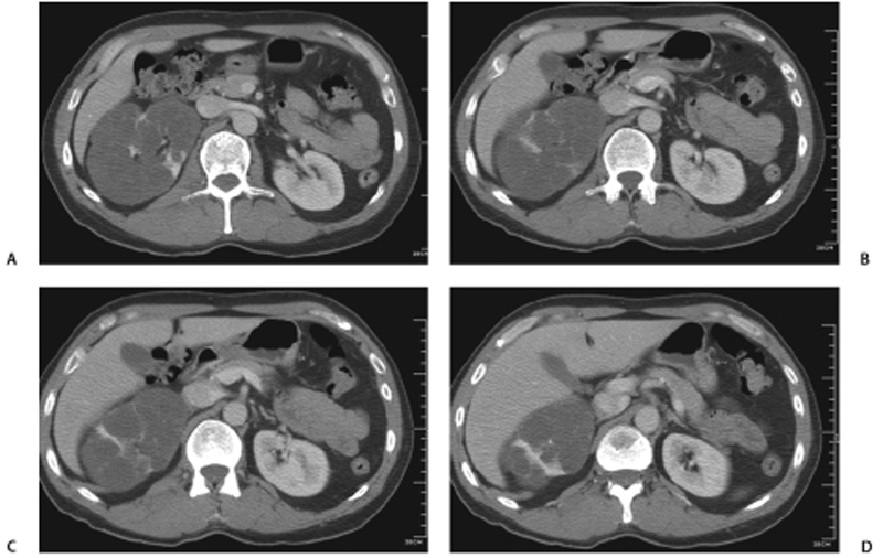
CASE 61 Incidental finding on an abdominal computed tomography (CT) scan. Fig. 61.1 (A–D) Select postcontrast CT images demonstrate multiple low-attenuation cysts throughout the right kidney, with intervening enhancing septations or renal parenchyma. No mural calcification or nodules are seen. The left kidney and other visualized abdominal viscera appear normal. Select computed tomography (CT) postcontrast images demonstrate multiple low-attenuation cysts throughout the right kidney, with intervening enhancing septations or renal parenchyma (Fig. 61.1). No mural calcification or nodules are seen. The left kidney and other visualized abdominal viscera appear normal. Localized cystic disease of the kidney
Clinical Presentation
Radiologic Findings
Diagnosis
Differential Diagnosis
Discussion
Background
Related posts:


Stay updated, free articles. Join our Telegram channel

Full access? Get Clinical Tree


