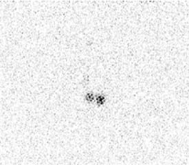
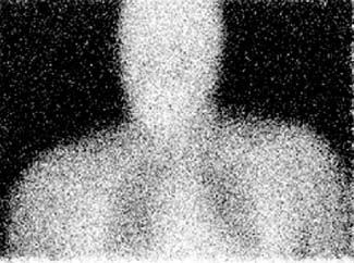
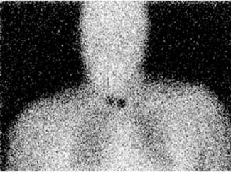
CASE 61 A 48-year-old woman with a history of thyroid cancer has undergone thyroidectomy and requires evaluation for thyroid remnant tissue and possible metastases. Fig. 61.1 Emission parallel-hole collimator view, 131I. Fig. 61.2 Transmission parallel-hole collimator view, 57Co. Fig. 61.3 Composite image: 131I + 57Co. • 5.0 mCi of 131I administered orally at 72 hours before scan • Use of a high-energy collimator for parallel-hole collimator imaging of the neck and thorax without (Fig. 61.1) and with (Fig. 61.2) cobalt 57 flood source behind patient (ie, patient between flood source and detector) • Emission image on 131I photopeak (364 keV); transmission image on 57Co photopeak (122 keV) without moving patient; superimposition of the two images (131I and 57Co) for composite image(Fig. 61.3) Figures 61.1, 61.2, and 61.3 all show a silhouette of the head, torso, and lungs, with two discrete foci in the thyroid bed. No other abnormal 131I-avid sites. • Residual functioning thyroid tissue in the thyroid bed • Surface contamination Remnants in thyroid bed only. No regional or distant metastases. Diagnosis final, no clinical follow-up.
Clinical Presentation
Technique
Image Interpretation
Differential Diagnosis
Diagnosis and Clinical Follow-Up
Discussion
Related posts:


Stay updated, free articles. Join our Telegram channel

Full access? Get Clinical Tree


