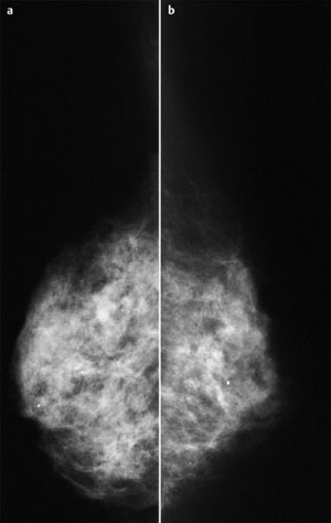
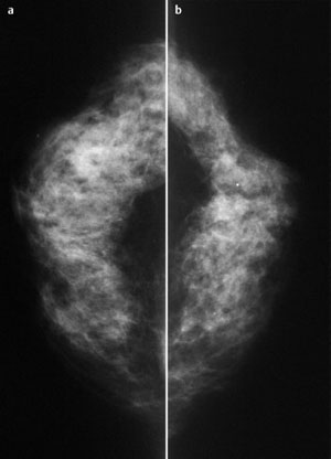
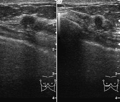
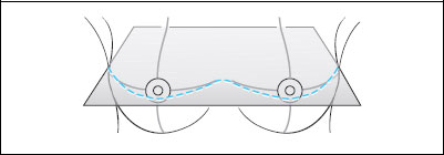
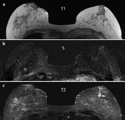
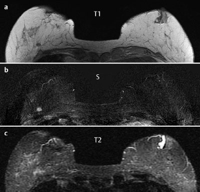
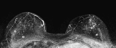
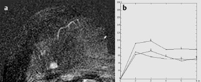
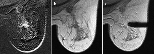
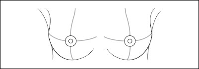
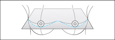
Case 62 Indication: Further investigation of suspicious finding in sonography of the left breast. History: Unremarkable. Risk profile: No increased risk. Age: 60 years. No findings. No findings. The suspicious lesion detected in the left breast in sonography conducted elsewhere was not reproducible. Fig. 62.1 a,b Conventional mammography, MLO view [imaging not performed by authors]. Fig. 62.2 a,b Conventional mammography, CC view [imaging not Performed by authors]. Fig. 62.3 Targeted repeat sonography in view of the MRI findings. Fig. 62.4 a-c Contrast-enhanced MRI of the breasts. Fig. 62.5 a-c Contrast-enhanced MRI of the breasts. Fig. 62.6 Contrast-enhanced MR mammography. Maximum intensity projection. Fig. 62.7 a,b Signal-to-time curves. Please characterize ultrasound, mammography, and MRI findings. What is your preliminary diagnosis? What are your next steps? Ultrasound showed inhomogeneous echogenicity of the tissue in both breasts with multiple hypoechoic, compressible lesions. The lesion between the upper quadrants of the left breast detected in a previous examination could not be reproduced. After MR mammography, further targeted sonography between the lower quadrants of the right breast showed a hypoechoic lesion (diameter 8 mm) with displacement of a Cooper’s ligament. There was partial distal shadowing. No relevant architectural distortions. US BI-RADS3. The glandular tissue was bilaterally symmetric and extremely dense, ACR type 4. Under these limiting conditions-even retrospectively-no suspicious masses or densities were visible, particularly between the lower quadrants of the right breast. There were no microcalcifications suspect for malignancy. BI-RADS right 1/left 1. PGMI: MLO view I (asymmetry, right pectoral muscle angle < 20°, left not depicted, inframammary fold incompletely depicted); CC view P. MRI demonstrated a lobulated, ill-defined, inhomogeneously* enhancing mass, 8 mm in diameter, between the lower quadrants of the right breast. The lesion had marked initial signal increase and postinitial washout as well as reduced signal in T2-weighted imaging. There were no suspicious findings in the upper quadrants of the left breast. In maximum intensity projection, additional enhancing foci with no criteria of malignancy were visible. There was also a duct ectasia on the left side behind the nipple. MRI Artifact Category: 1 MRI Density Type: 1
Clinical Findings
Ultrasound (not shown)

Ultrasound
Mammography
MR Mammography
MRM score | Finding | Points |
Shape | lobulated | 0 |
Border | ill-defined | 1 |
CM Distribution | inhomogeneous | 1 |
Initial Signal Intensity Increase | strong | 2 |
Post-initial Signal Intensity Character | wash-out | 2 |
MRI score (points) |
| 6 |
MRI BI-RADS |
| 5 |
 Differential Diagnosis
Right breast: Carcinoma (medullary?), adenosis, fibroadenoma, papilloma.
* Differentiation between inhomogeneity and internal septations was not possible.
Clinical Findings | right 1 | left 1 |
Ultrasound | right 3 | left 1 |
Mammography | right 1 | left 1 |
MR Mammography | right 5 | left 1 |
BI-RADS Total | right 5 | left 1 |
Procedure
MR-guided vacuum biopsy (Fig. 62.8), since the correlation of the findings in MRI and in ultrasound was not firmly established.
Histopathology
Invasive ductal carcinoma.
Further procedure
Tumor excision following preoperative MR-guided localization.
Fig. 62.8 a-e MR-guided vacuum biopsy of the right breast.
Fig. 62.9 a-c MR-guided localization of the lesion in the right breast.
Diagnosis
IDC pTIb, pNO, G2
Stay updated, free articles. Join our Telegram channel

Full access? Get Clinical Tree


