Case 62

 Clinical Presentation
A 64-year-old man with flank pain.
 Imaging Findings

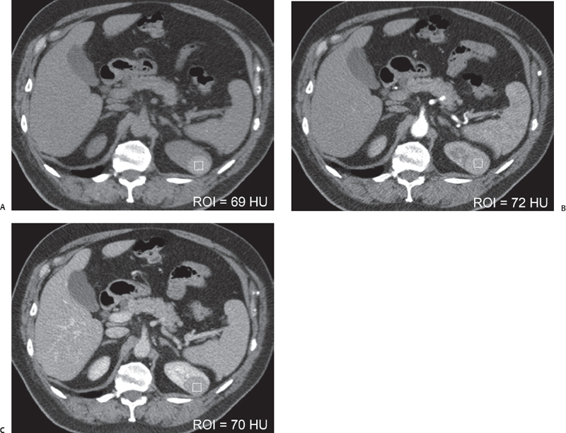
(A) Precontrast axial computed tomography (CT) image at the level of the upper poles of the kidneys shows a focal lesion (arrow) with homogeneously high attenuation measured at 69 Hounsfield units (HU). The margins of the lesion are smooth and sharply defined. There is no calcification. No perinephric stranding is seen. (B) Contrast-enhanced corticomedullary phase axial CT image at the same level as in Figure A shows the attenuation value of the lesion (arrow) at 72 HU, indicating no enhancement during this phase. (C) Contrast-enhanced nephrographic phase axial CT image at the same level as in Figures A and B shows the attenuation value of the lesion (arrow) at 70 HU, indicating no enhancement during this phase.
 Differential Diagnosis
• Hemorrhagic renal cyst: A round lesion that has sharply marginated smooth walls and homogeneously high attenuation with no enhancement is characteristic.
• Renal neoplasm: A renal lesion that does not have fluid signal intensity raises the question of a renal neoplasm. However, the lack of enhancement after intravenous contrast shows that there is no solid tissue within this lesion. Therefore, this lesion is unlikely to be a renal neoplasm.
Stay updated, free articles. Join our Telegram channel

Full access? Get Clinical Tree


