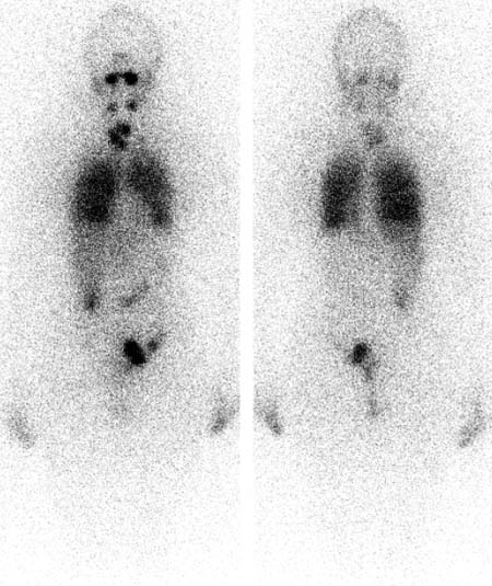
CASE 62 A 42-year-old woman with stage I papillary thyroid cancer presents for pre-therapy whole-body scan, 131I ablation therapy, and post-therapy whole-body scan. Her serum thyroglobulin level is markedly elevated at 300 ng/mL (normal, < 0.5 ng/mL). Fig. 62.1 Whole-body parallel-hole collimator views, anterior and posterior projections, 123I (pre-therapy, diagnostic dose). Fig. 62.2 Whole-body parallel-hole collimator views, anterior and posterior projections, 131I (post-therapy dose). • Pre-therapy scan: 2 mCi of oral 123I 24 hours before scan • Post-therapy scan: 200 mCi of oral 131I one week before scan • Whole-body diagnostic (pre-therapy) imaging (Fig. 62.1) • Whole-body post-therapy imaging (Fig. 62.2) On the diagnostic (pre-therapy) 123I scan, multiple small, scattered foci in the neck suggest remnant tissue and possibly regional nodal metastases. On the post-therapy scan, marked diffuse activity throughout the lungs and liver is new; the neck activity is much more intense and extensive. • Metastatic thyroid carcinoma • Salivary or urinary contamination Thyroid remnant tissue, regional lymph node metastases, and diffuse pulmonary metastases; physiologic uptake of tracer in the liver on post-therapy scan. Diagnosis final.
Clinical Presentation
Technique
Image Interpretation
Differential Diagnosis
Diagnosis and Clinical Follow-Up
Discussion
Related posts:


Stay updated, free articles. Join our Telegram channel

Full access? Get Clinical Tree


