Case 63
 Clinical Presentation
A 43-year-old woman with weight loss and a flank mass.
 Imaging Findings
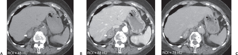
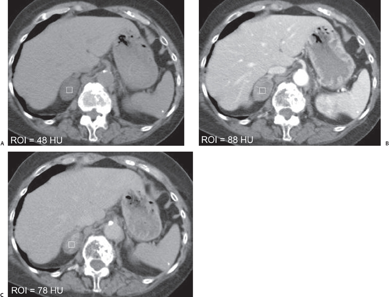
(A) Non–contrast-enhanced axial computed tomography (CT) image at the level of the adrenal glands shows that the right adrenal gland has been replaced by a mass (arrow) with a heterogeneous appearance due to areas of high and low attenuation. The attenuation has been measured at 48 Hounsfield units (HU). (B) Contrast-enhanced axial CT image at the same level as in Figure A obtained 60 seconds after the initiation of a contrast injection shows heterogeneous enhancement of the right adrenal mass (arrow). The attenuation has been measured at 88 HU. (C) Contrast-enhanced axial CT image at the same level as in Figures A and B obtained after a 10-minute delay shows persistence of the heterogeneous enhancement in the right adrenal mass (arrow). The attenuation has been measured at 78 HU.
Stay updated, free articles. Join our Telegram channel

Full access? Get Clinical Tree


