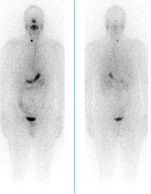
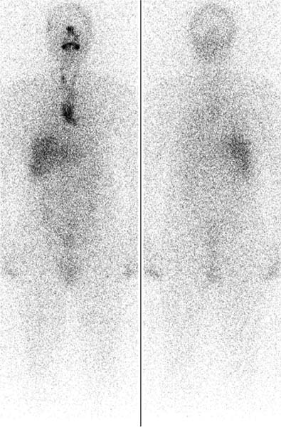
CASE 63 A 34-year-old man with papillary thyroid cancer has undergone thyroidectomy and presents for imaging after radioiodine ablation. Fig. 63.1 Whole-body parallel-hole collimator views, anterior and posterior projections, 123I (pre-therapy). Fig. 63.2 Whole-body parallel-hole collimator views, anterior and posterior projections, 131I (therapeutic dose). • Pre-therapy scan: 2 mCi of 123I administered orally for diagnostic scan 2 days before 131I therapy • Post-therapy scan: 100 mCi of therapeutic 131I administered orally 1 week before scan • Whole-body 123I imaging in anterior and posterior projections with a dual-detector gamma camera equipped with low-energy collimators (Fig. 63.1) • Whole-body 131I imaging in anterior and posterior projections with a dual-detector gamma camera equipped with high-energy collimators (Fig. 63.2) Physiologic distribution and a focus of thyroid tissue in the right side of the neck are seen on pre-therapy diagnostic 123I imaging (Fig. 63.1
Clinical Presentation
Technique
Image Interpretation
![]()
Stay updated, free articles. Join our Telegram channel


Full access? Get Clinical Tree


