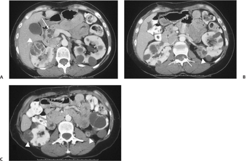
CASE 63 Routine follow-up study in an asymptomatic patient with known von Hippel-Lindau disease. Fig. 63.1 (A) Postcontrast CT image shows bilateral renal cysts. There is a large cyst within the right kidney with thick enhancing septations (arrowhead). Note the multiple pancreatic cysts (arrow). (B) Axial image shows a mixed solid and cystic lesion in the left kidney (arrowhead), along with an enhancing nodule in the chord (arrow). (C)An additional complex cyst with enhancing septation is seen in the right kidney (arrowhead), along with a small enhancing lesion in the left kidney (arrowhead). Postcontrast computed tomography (CT) images demonstrate bilateral multiple low-attenuation renal cysts, some of which show enhancing nodules or septations or have solid components (Fig. 63.1). There is also an enhancing lesion within the spinal cord, as well as multiple cysts seen in the pancreatic head. Von Hippel-Lindau disease
Clinical Presentation
Radiologic Findings
Diagnosis
Differential Diagnosis
Discussion
Related posts:


Stay updated, free articles. Join our Telegram channel

Full access? Get Clinical Tree


