Case 64
Indication: Monitoring after bilateral breast cancer.
History: Invasive lobular carcinoma of the left breast 5 years previously and ductal carcinoma in situ of the right breast 3 years previously. Bilateral breast conservation therapy and radiation therapy.
Risk profile: Increased by earlier bilateral cancer.
Age: 52 years.
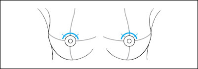
Clinical Findings
Normal scarring of both breasts. No palpable findings.
Ultrasound (not shown)
Bilateral lumpectomy scars; no other unusual findings.
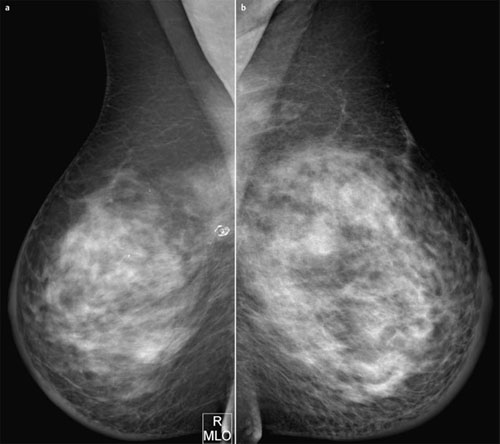
Fig. 64.1 a,b Digital mammography, MLO view.
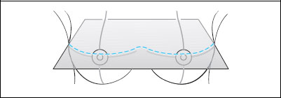
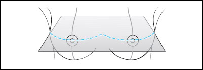
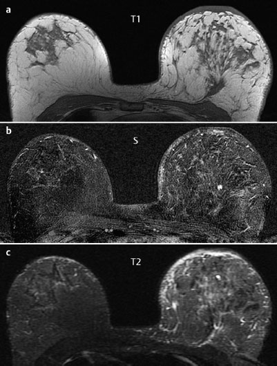
Fig. 64.2 a,b Contrast-enhanced MRI of the breasts.
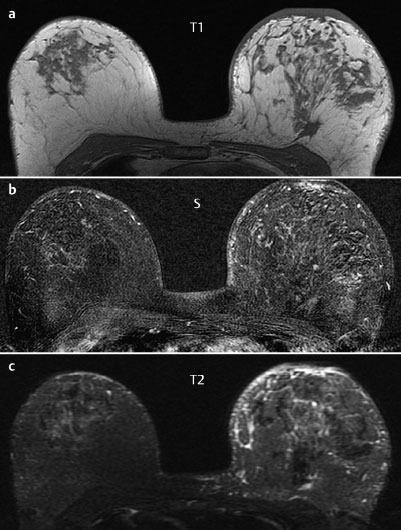
Fig. 64.3 a,b Contrast-enhanced MRI of the breasts.
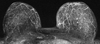
Fig. 64.4 a,b Contrast-enhanced MR mammography. Maximum intensity projection.
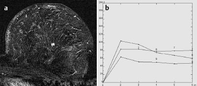
Fig. 64.5 a,b Signal-to-time curves.

|
Please characterize mammography and MRI findings.
What is your preliminary diagnosis?
What are your next steps? |
This case presents the routine follow-up imaging of a 52-year-old woman after bilateral breast cancer 3 and 5 years previously.
Ultrasound
Bilateral hypoechoic lumpectomy scars were visible. There were no suspicious findings. US BI-RADS right 2/left 2 (scars). (Not shown).
MR Mammography
Tl-weighted MRI demonstrated a postoperative architectural distortion in the center of the left breast. Within this area there was an ill-defined lesion 6 mm in diameter that showed postcontrast hypervascularization, initial signal increase of 110%, postinitial washout, and reduced signal in T2-weighted imaging.
Mammography
Mammograms showed bilaterally symmetric, inhomogeneously dense parenchyma, ACR type 3. Skin thickening due to previous treatment was visible around the circumference of both breasts, as well as postoperative architectural distortion in the upper quadrants of the right breast toward the chest wall. Here there were also eggshell-like calcifications, suggesting an oil cyst. There were no suspicious lesions or microcalcifications. BI-RADS right 2/left 1. PGMI: MLO view G (inframammary fold).
MRI Artifact Category: 1
MRI Density Type: 2
 Differential Diagnosis


