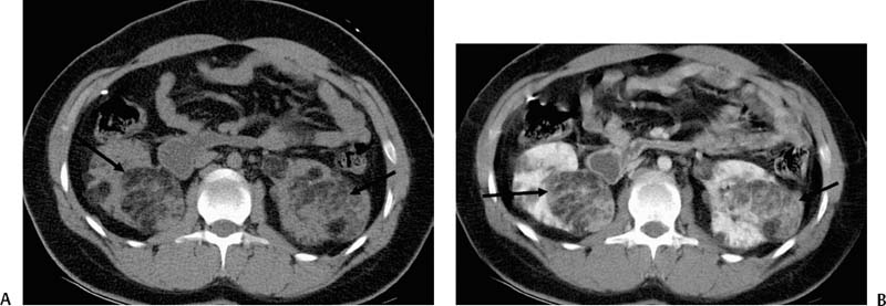
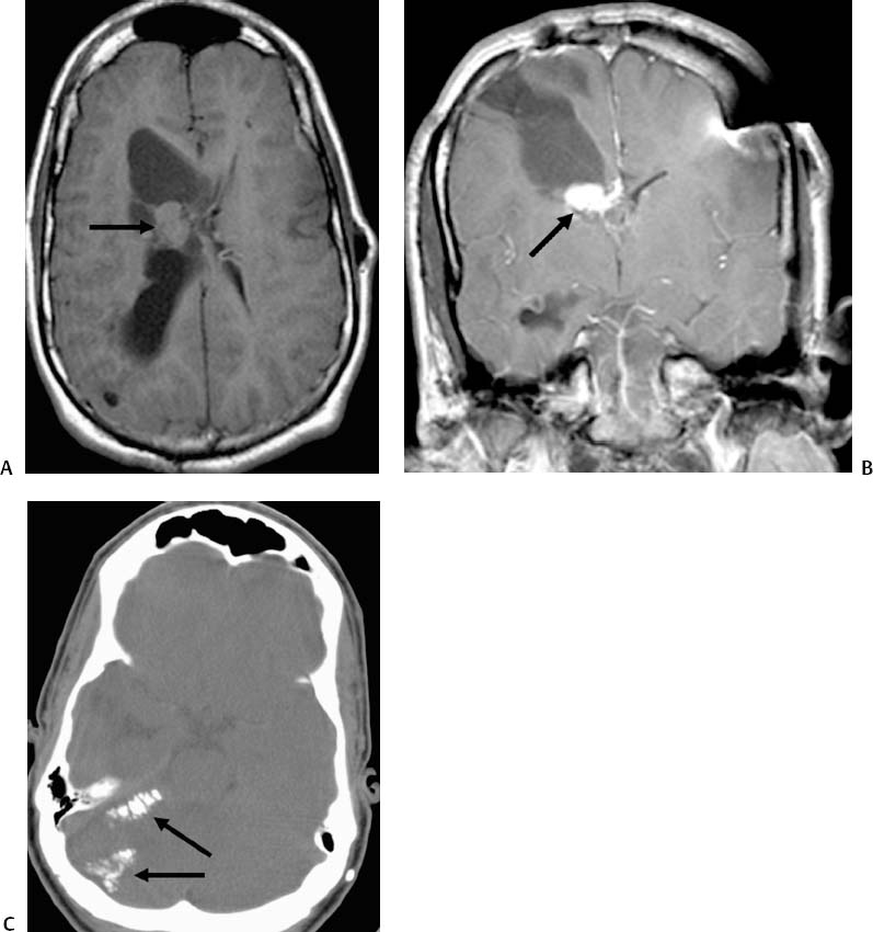
CASE 64 An 18-year-old male presented with left-sided flank pain and gross hematuria. Figure 64-1 CT of the abdomen. (A) Precontrast-enhanced and (B) postcontrast-enhanced CT images of the abdomen show masses (arrows) in both kidneys. Note the irregular enhancement. Low-density components of these masses represent fat elements. Contrast-enhanced CT scans show bilateral renal masses (Figs. 64-1A, B). The masses were heterogenous in appearance and contained fat. Brain imaging of this patient obtained previously shows the presence of an enhancing mass lesion in the region of the enlarged right lateral ventricle (Figs. 64-2A, B). Coarse calcifications are present in the brain on CT (Fig. 64-2C). Bleeding angiomyolipoma in a patient with tuberous sclerosis (TS). The right common femoral artery was catheterized using the Seldinger technique, and a 5-French (F) sheath was inserted. An aortogram was obtained using a 5F pigtail catheter (Boston Scientific, Natick, Massachusetts), which revealed bilateral vascular lesions in the kidneys (Figs. 64-3A, B). The right renal artery was selectively catheterized with a 5F RC-1 catheter (Boston Scientific, Natick, Massachusetts), and renal arteriography was performed (Fig. 64-3C
Clinical Presentation
Radiologic Studies
Cross-Sectional Imaging
Diagnosis
Treatment
Equipment
Selective Embolization
![]()
Stay updated, free articles. Join our Telegram channel


Full access? Get Clinical Tree


