CASE 64 A 43-year-old woman presents with a recent diagnosis of renal stones. The patient has elevated calcium and parathyroid hormone levels. Fig. 64.1 Top row: 20-minute images; bottom row: 2-hour images. • Approximately 20 mCi of 99mTc-sestamibi injected intravenously • High-resolution collimator • Anterior view of the neck and chest done at 20 minutes and 2 hours after injection • Pinhole view of the thyroid bed done at 20 minutes and 2 hours after injection Initial images at 20 minutes (Fig. 64.1, top row) show relatively diffuse uptake in the thyroid and a focus of increased tracer uptake in the right lower pole. Two-hour–delayed images (Fig. 64.1, bottom row) show washout of tracer from the thyroid and residual tracer concentration in the region of the right lower pole. • Parathyroid adenoma • Parathyroid hyperplasia • Thyroid adenoma (less common) • Thyroid carcinoma • Parathyroid carcinoma Ultrasound demonstrated a possible nodule on the left side, but no abnormality on the right side. Surgical exploration revealed a 3 × 2-cm right inferior parathyroid adenoma. Parathyroid hormone levels returned to normal after resection of the adenoma.
Clinical Presentation
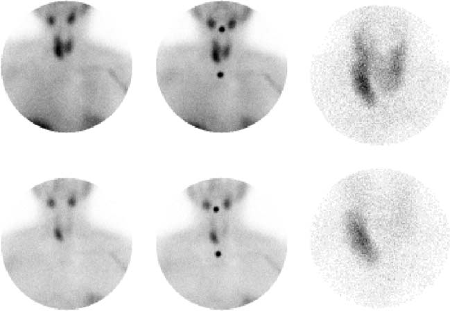
Technique
Image Interpretation
Differential Diagnosis
Diagnosis and Clinical Follow-Up
Discussion
Related posts:


Stay updated, free articles. Join our Telegram channel

Full access? Get Clinical Tree


