Case 65
 Clinical Presentation
A 43-year-old man with left flank pain.
 Imaging Findings
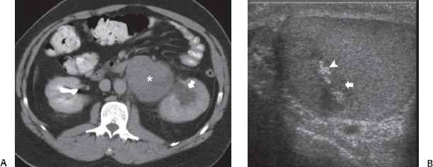
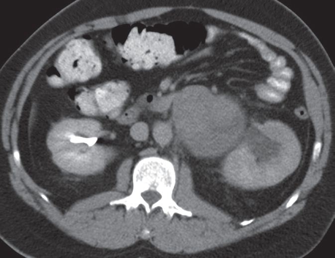
(A) Contrast-enhanced excretory computed tomography image of the abdomen at the level of the kidneys show a lobulated mass (asterisk) in the left para-aortic region at the level of the left renal hilum. There is mild hydronephrosis (arrow) of the left kidney with lack of excretion of contrast in comparison with the right kidney. (B) Sonogram of the left testis shows an ill-defined focal lesion (arrow) with calcifications (arrowhead).
 Differential Diagnosis
• Retroperitoneal lymphadenopathy due to metastases from a possible testicular cancer:
Stay updated, free articles. Join our Telegram channel

Full access? Get Clinical Tree


