CASE 65
Clinical Presentation
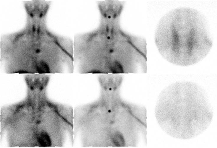
A 34-year-old woman presents with a ureteral stone. Subsequent workup reveals elevated calcium and parathyroid hormone levels.

Fig. 65.1 Top row: 20-minute images; bottom row: 2-hour images. The middle column shows marker views.
Technique
• Approximately 20 mCi of 99mTc-sestamibi injected intravenously
• High-resolution collimator
• Anterior view of the neck and chest done at 20 minutes and 2 hours after injection
• Pinhole view of the thyroid bed done at 20 minutes and 2 hours after injection
Image Interpretation


