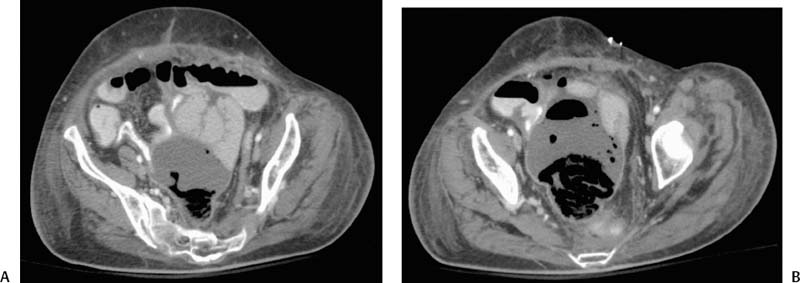
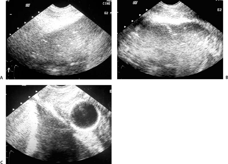
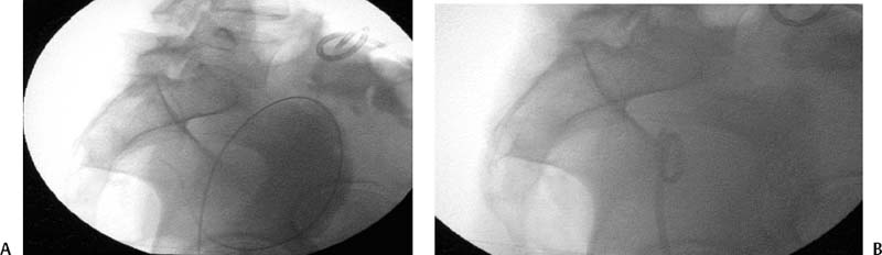
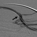
CASE 65 A 62-year-old female presented with fever, leukocytosis, and abdominal pain 2 weeks after undergoing low perineal resection for rectal carcinoma. Figure 65-1A and B Contrast-enhanced CT of the pelvis (A). Scans at two different levels showed a fluid collection in the peluic cul-de-sac (B). Folded radiolucent material in the posterior aspect of the collection represents surgical foam intentionally left in the surgical bed for hemostasis. Contrast-enhanced CT shows a pelvic fluid collection (Fig. 65-1) containing radiolucent material representing surgical foam, intentionally left in the operative bed for control of bleeding. Pelvic abscess. The patient was placed in the lateral decubitus position on the angiographic table. A transrectal ultrasound probe was inserted and used to localize the abscess (Fig. 65-2). The abscess was punctured with an 18-gauge trocar needle, the inner stylet of the needle was removed, and a 0.035-inch Amplatz wire (Boston Scientific, Natick, MA) was advanced through the needle into the fluid collection (Fig. 65-3A). Once access with the wire was secured, the needle and ultrasound probe were removed over the wire. Dilatation of the tract was performed, a 10-French (F) catheter was advanced into the abscess over the wire, and the wire was removed (Fig. 65-3B). The catheter was secured with tape to the skin and placed to bulb suction. Follow-up CT showed near-complete resolution (Fig. 65-4). Figure 65-3 (A) A wire was advanced into the fluid collection. (B) A 10F pigtail catheter was placed over the wire.
Clinical Presentation
Radiologic Studies
Diagnosis
Treatment
Equipment
Related posts:


Stay updated, free articles. Join our Telegram channel

Full access? Get Clinical Tree


