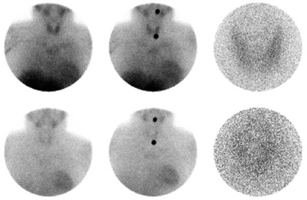
CASE 66 A 34-year-old man presents with a long history of hyperparathyroidism. A sestamibi study is obtained for parathyroid localization. Fig. 66.1 Top row: 20-minute images; bottom row: 2-hour images. The middle column shows marker views. • Approximately 20 mCi of 99mTc-sestamibi injected intravenously • High-resolution collimator • Anterior view of the neck and chest done at 20 minutes and 2 hours after injection • Pinhole view of the thyroid bed done at 20 minutes and 2 hours after injection Normal uptake is seen in the region of the thyroid bed (Fig. 66.1). A vague blush in the region of the mediastinum is also noted but not seen clearly enough to be diagnostic. Retention of nuclide in this area is unchanged on the delayed images, which is consistent with delayed washout. (For mediastinal uptake)
Clinical Presentation
Technique
Image Interpretation
Differential Diagnosis
Stay updated, free articles. Join our Telegram channel

Full access? Get Clinical Tree


