
MRM score | Finding | Points |
Shape | irregular/segmental | 1 |
Border | ill-defined | 1 |
CM Distribution | inhomogeneous | 1 |
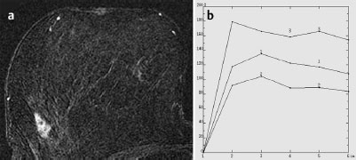
Initial Signal Intensity Increase | strong | 2 |
Post-initial Signal Intensity Character | wash-out | 2 |
MRI score (points) |
| 7 |
MRI BI-RADS |
| 5 |
 Differential Diagnosis
Local tumor relapse, focal (e.g., granulomatous) mastitis, focal adenosis.
BI-RADS Categorization | ||
Clinical Findings | right 1 | left 1 |
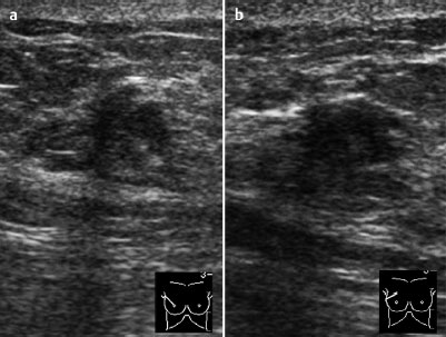
Ultrasound | right 3 | left 1 |
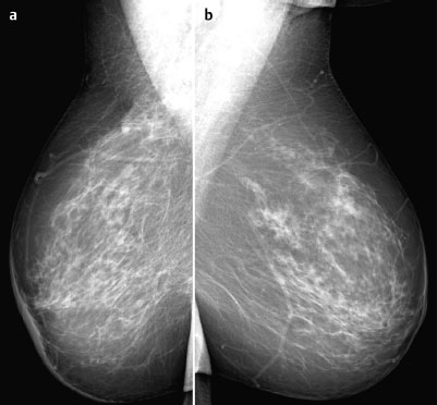
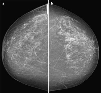
Mammography | right 3 | left 1 |
MR Mammography | right 5 | left 1 |
BI-RADS Total | right 5 | left 1 |
Procedure
MR-guided vacuum biopsy of the lesion in the upper outer quadrant of the right breast.
Histology
Invasive ductal carcinoma.
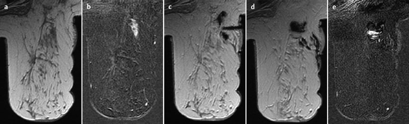
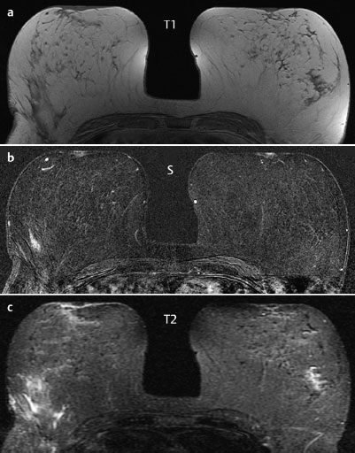
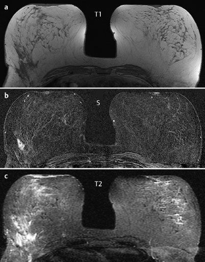
Fig.67.8 a-e MR-guided vacuum biopsy of the right breast.
a Precontrast TI-weighted image.
b Reproducibility of the segmental enhancement in subtraction image.
c Coaxial needle after local anesthesia.
d Findings after biopsy in precontrast TI-weighted image.
e Subtraction image after further contrast administration with enhancement due to bleeding. No residual enhancing tumor component.
Diagnosis
Local tumor relapse after breast conservation therapy.
Stay updated, free articles. Join our Telegram channel

Full access? Get Clinical Tree


