Case 68
 Clinical Findings
A 51-year-old woman with recurrent urinary tract infection.
 Imaging Findings
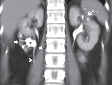
Maximum intensity projection image of a computed tomography (CT) urogram shows mild dilatation of the collecting system of the lower half of the right kidney (asterisk) with severe parenchymal scarring (thick arrow). Two ureters (arrowheads) are seen draining the right kidney. The upper half of the right kidney is normal. Some scarring is seen at the upper pole of the left kidney (thin arrow).
 Differential Diagnosis
• Duplex right renal collecting system with scarring of the lower moiety due to reflux: Chronic hydronephrosis of the left upper pole with a normal lower pole is suggestive of a lesion affecting the upper pole calices. The presence of a nonopacified dilated additional ureter adjacent to the opacified normal left ureter further supports the diagnosis.
• Infundibular stricture of the right lower pole infundibulum: This would cause chronic hydronephrosis of the right lower pole calices with overlying parenchymal atrophy. However, in this case, the parenchymal atrophy and scarring are out of proportion to the mild degree of collecting system dilatation.
• Urothelial neoplasm of the lower right infundibulum:
Stay updated, free articles. Join our Telegram channel

Full access? Get Clinical Tree


