Case 69
 Clinical Presentation
A 43-year-old woman with hematuria.
 Imaging Findings
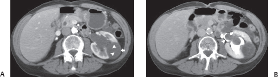
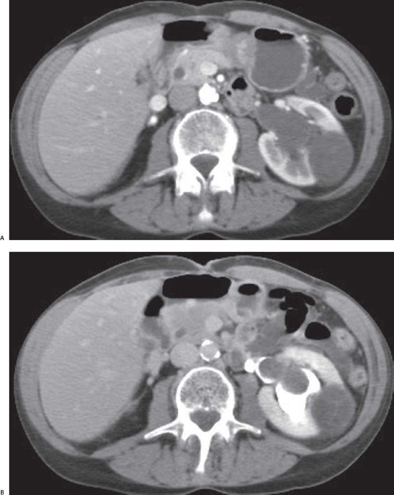
(A) Contrast-enhanced computed tomography (CT) image in nephrographic phase at the level of the kidneys shows a cystic-appearing lesion (arrow) within the left kidney. There is no obvious enhancing soft tissue. Fine septa (arrowheads) are visible in the lesion. (B) Contrast-enhanced CT in excretory phase at a level lower than that in Figure A shows herniation of the cystic lesion (arrow) into the opacified renal pelvis (arrowhead).
 Differential Diagnosis
• Multilocular cystic nephroma (MLCN): A cystic mass with multiple thin septa and herniation into the renal pelvis is characteristic.
• Cystic renal cell carcinoma:
Stay updated, free articles. Join our Telegram channel

Full access? Get Clinical Tree


