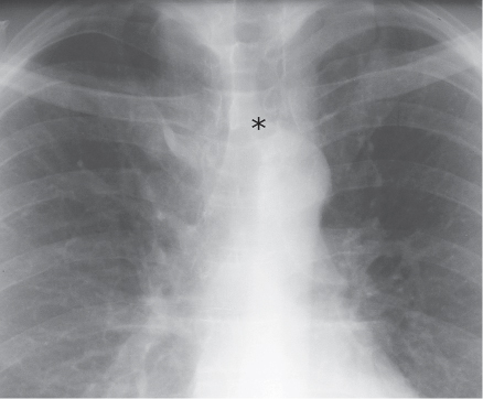
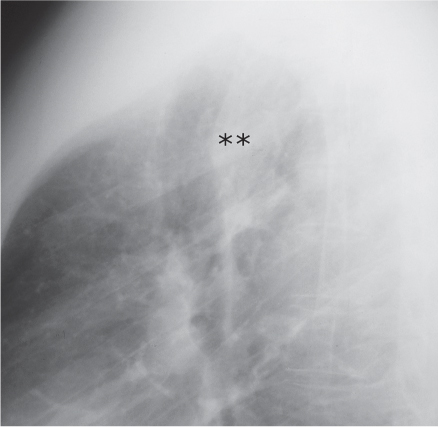
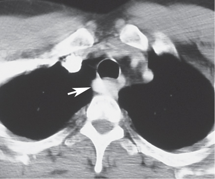
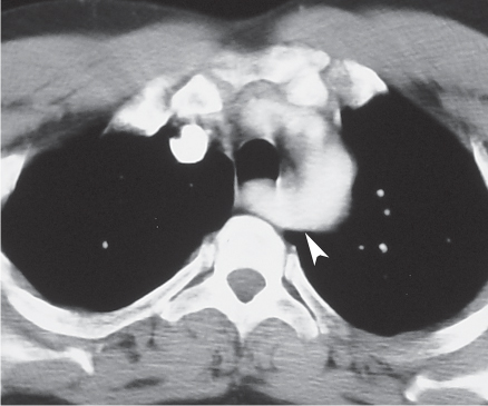
CASE 7 Asymptomatic 42-year-old man Coned-down PA (Fig. 7.1) and lateral (Fig. 7.2) chest radiographs demonstrate a left aortic arch and a tubular opacity (asterisk) that arises from its superior aspect, projects over the tracheal lumen, and courses obliquely toward the right upper extremity (Fig. 7.1). Note the presence of an accessory azygos fissure. The lateral radiograph (Fig. 7.2) demonstrates a soft-tissue opacity (double asterisk) that produces mass effect on the dorsal trachea. Contrast-enhanced chest CT (mediastinal window) (Figs. 7.3, 7.4) demonstrates an aberrant right subclavian artery (arrow) (Fig. 7.3) that arises from a diverticulum of Kommerell (arrowhead) (Fig. 7.4). Aberrant Right Subclavian Artery None Fig. 7.1 Fig. 7.2 Fig. 7.3 Fig. 7.4 (Images 7.1–7.4 are courtesy of Maysiang Lesar, MD, National Naval Medical Center, Bethesda, Maryland.) An aberrant right subclavian artery arises as the last branch off a left-sided aortic arch and courses obliquely and superiorly behind the trachea and esophagus to resume its normal course. It is typically an isolated anomaly, represents one of the most common aortic arch branching anomalies, and affects up to 0.5% of the population, with a reported prevalence of 0.4–2%.
 Clinical Presentation
 Radiologic Findings
 Diagnosis
 Differential Diagnosis
 Discussion
Background
Etiology
Related posts:
![]()
Stay updated, free articles. Join our Telegram channel

Full access? Get Clinical Tree


Radiology Key
Fastest Radiology Insight Engine


