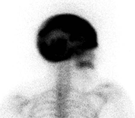
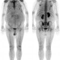
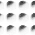
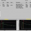
CASE 7 An 82-year-old woman presents with a recent history of bilateral hearing loss (Figs. 7.1 and 7.2). Fig. 7.1 Fig. 7.2 • A 20 mCi dose of 99mTc-HDP is administered intravenously. • Whole-body and spot images are obtained 3 hours after tracer administration. • Emphasize the importance of oral hydration to improve soft tissue and bladder clearance. Anterior whole-body (Fig. 7.1) and lateral skull (Fig. 7.2) views show intense, abnormal uptake within the calvarium, with relatively faint visualization of the remainder of the axial skeleton and the appendicular portions of the skeleton. The injection site is seen in the left hand.
Clinical Presentation
Technique
Image Interpretation
Stay updated, free articles. Join our Telegram channel

Full access? Get Clinical Tree


