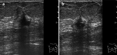
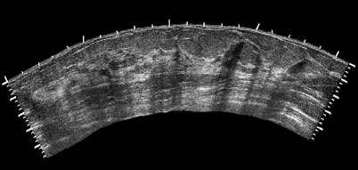
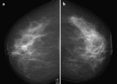
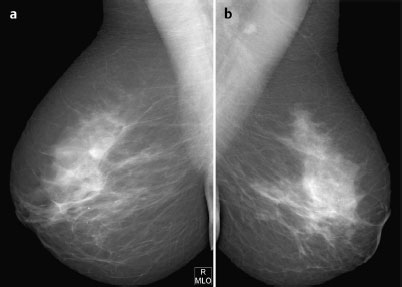
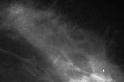
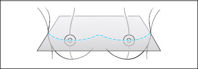
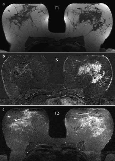
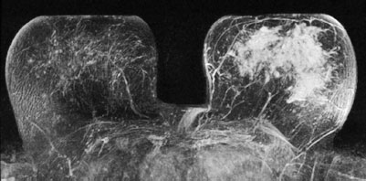
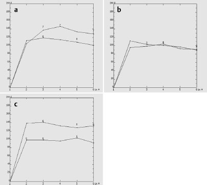
Case 71 Indication: Screening. Mastodynia of the left breast. History: Unremarkable. Risk profile: No increased risk. Age: 67 years. Normal. Fig. 71.1 a, b Sonography. Fig. 71.2 Sonography. Panoramic view of the left breast. Fig. 71.3a,b Digital mammography, CC view. Fig. 71.4a,b Digital mammography, MLO view. Fig. 71.5 Magnification view of the outer quadrants of the left breast (CC). Fig. 71.6a–c Contrast-enhanced MRI of the breasts Fig. 71.7 Contrast-enhanced MR mammography. Maximum intensity projection. Fig. 71.8a–c Signal-to-time curves of representative regions of the left breast. Please characterize ultrasound, mammography, and MRI findings. What is your preliminary diagnosis? What are your next steps? There was a highly suspicious irregular lesion in the left breast with the long axis perpendicular to the skin and with hyperechoic margin, distortion of ligamental structures, and pathological distal echo shadowing. Panoramic view of the left breast demonstrated multiple linear distal shadows that, taken together with the primary lesion, also point to malignancy. There was a small cyst in the right breast. US BI-RADS right 2/left 5. The parenchymal texture was inhomogeneously dense, ACR type 3, and asymmetric (right>left). In the upper outer quadrant of the left breast, magnification view depicted multiple predominantly amorphous microcalcifications (Fig. 71.5). There were no circumscribed lesions in the left breast. A small round lesion was visible in the right breast (consistent with the cyst seen in ultra sound). BI-RADS right 2/left 5. PGMICC view P; MLO view G (inframammary fold). Precontrast images of both breasts were unremarkable. However, postcontrast imaging demonstrated unilateral diffuse enhancement of the entire nonlipomatous tissue of the left breast. Signal-time curves were characteristic of malignancy. MRI Artifact Category: 1 MRI Density Type: 1
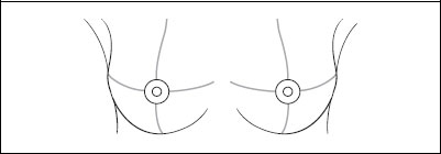
Clinical Findings

Ultrasound
Mammography
MR Mammography
MRM score | Finding | Points |
Shape | irregular | 1 |
Border | ill-defined | 1 |
CM Distribution | homogeneous | 0 |
Initial Signal Intensity Increase | strong | 2 |
Post-initial Signal Intensity Character | wash-out | 2 |
MRI score (points) |
| 6 |
MRI BI-RADS |
| 5 |
 Preliminary Diagnosis
Carcinoma of the left breast (most likely diffuse invasive lobular carcinoma, although the calcifications are not typical for this). Non-mass lesion seen in MRI.
 Differential Diagnosis
Mastitis (very low probability).
BI-RADS Categorization | ||
Clinical Findings | right 1 | left 1 |
Ultrasound | right 2 | left 5 |
Mammography | right 2 | left 5 |
MR Mammography | right 1 | left 5 |
BI-RADS Total | right 2 | left 5 |
Procedure
Histological analysis of the suspicious finding in the left breast with US-guided core biopsy.
Histology of the left breast
Ductal carcinoma in situ (DCIS) accompanied by atypical ductal hyperplasia (ADH).
Histology
Extensive malignant tumor with multifocal micropapillary, low-grade ductal carcinoma in situ. Transformation into invasive ductal carcinoma in the upper outer quadrant of the left breast.
IDC pT1c+DCIS (multifocal), pN2a (4/12), G2.
Stay updated, free articles. Join our Telegram channel

Full access? Get Clinical Tree


