Case 71
 Clinical Presentation
A 24-year-old woman with menstrual irregularity.
 Imaging Findings
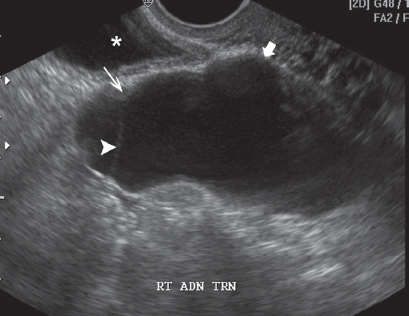
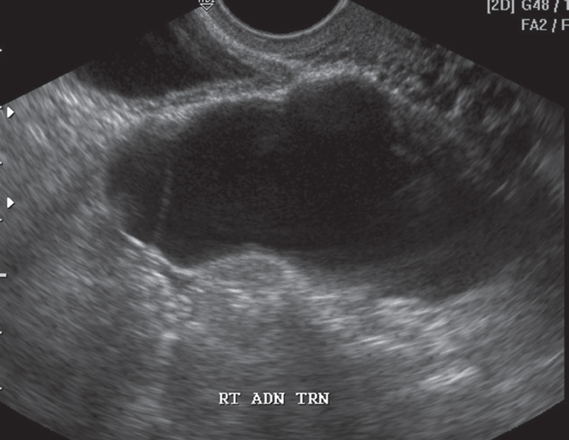
Pelvic ultrasound image of the right adnexa shows a fluid-filled tubular structure (short arrow) with thin walls lying posterior to the urinary bladder (asterisk). The contents are clear. There is a thin, incomplete septation (arrowhead) in the lesion with a subtle waist (long arrow) on the wall opposite the septation. No free peritoneal fluid is seen.
 Differential Diagnosis
• Hydrosalpinx: The tubular shape, incomplete septation, and waist on the opposite wall are characteristic of a dilated fallopian tube. The contents are clear, and there are no obvious signs of inflammation (wall edema, free fluid).
• Tubo-ovarian abscess:
Stay updated, free articles. Join our Telegram channel

Full access? Get Clinical Tree


