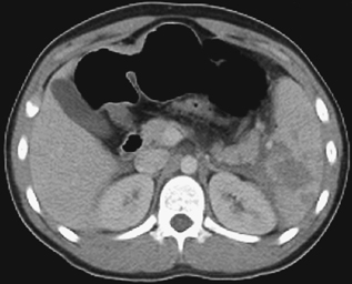
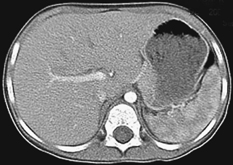
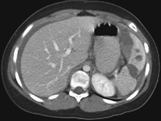
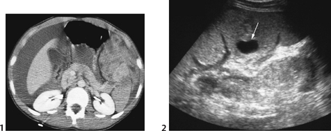
CASE 71 A 13-year-old boy presents with left chest pain following a snowboarding injury. Figure 71A Figure 71B Selected abdominal CT images demonstrate hypodensity of the spleen in its mid (Fig. 71A) and lower portions (Fig. 71B) without fluid in the lateral paracolic gutter. Fluid is seen tracking medially along the pancreatic tail. Figure 71C Normal CT pattern of early splenic enhancement following intravascular contrast administration. Splenic trauma The following are the mimickers: Other causes of hypodense lesions in the spleen: The spleen is commonly injured in children following blunt abdominal trauma, including motor vehicle accidents or child abuse. An underlying splenomegaly, which can occur, for example, in infectious mononucleosis, Gaucher’s disease, or splenic epidermoid cyst, is a risk factor. The past decades have seen confirmation of nonoperative management, avoiding unnecessary laparotomy and the risk of postsplenectomy infection. Trauma to the left side of the abdomen is often associated with injury to the spleen and left lung contusion or laceration. Rib fractures are less commonly seen in children than in adults because of the greater pliability of the child’s chest wall. Trauma to the left hemiabdomen is often associated with both splenic and renal trauma. Figure 71D Delayed splenic rupture. Patient presented with acute abdominal pain and syncope. There was a history of skiing accident 4 weeks prior. CT demonstrates hemoperitoneum, subcapsular splenic hematoma, and splenic fracture. Figure 71E (1) Traumatic pseudoaneurysm of splenic artery. Initial CT of teenage boy after roller-blading injury shows splenic laceration, perisplenic hyperdense clots, and evidence of extensive hemoperitoneum. No contrast extravasation was seen on initial scans. (2
Clinical Presentation

Radiologic Findings
Diagnosis
Differential Diagnosis
Discussion
Background
Etiology
![]()
Stay updated, free articles. Join our Telegram channel


Full access? Get Clinical Tree


