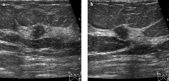
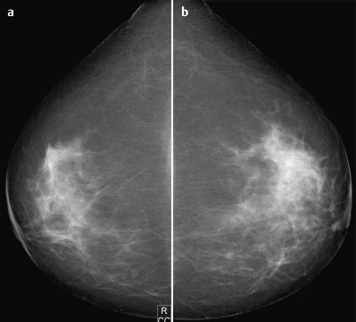
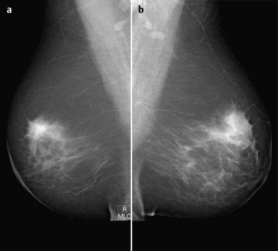
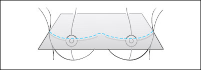
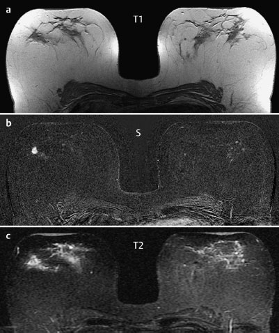
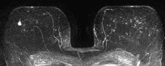
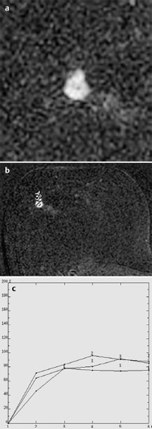
Case 72 Indication: Screening. History: Unremarkable. Risk profile: No increased risk. Age: 57 years. Fig. 72.1 a,b Ultrasound. Normal. Fig. 72.2a,b Digital mammography, CC view. Fig. 72.3a,b Digital mammography, MLO view. Fig. 72.4a–c Contrast-enhanced MRI of the breasts. Fig. 72.5 Contrast-enhanced MR mammography. Maximum intensity projection. Fig. 72.6a-c Enlarged view of lesion; signal-to-time curves. Please characterize ultrasound, mammography, and MRI findings. What is your preliminary diagnosis? What are your next steps? This case demonstrates the imaging studies of an asymptomatic woman presenting for screening. In the upper outer quadrant of the right breast, there was an ill-defined, lobulated, hypoechoic area within the glandular tissue with inhomogeneous internal texture. There was no echo alteration of the surrounding structures, but ligamental structures were clearly displaced by this region. US BI-RADS right 3. Mammograms showed inhomogeneously dense glandular tissue of ACR type 3 with a mild asymmetry of the upper outer quadrants. In the right breast there was a slight shrinking with some peripheral spiculation in the upper outer quadrant. There were no densities or definite lesions. No architectural distortions, and no microcalcifications. BI-RADS right 3/left 1. PGMI CC view P; MLO view G (inframammary fold). MRI demonstrated a single hypervascularized, ill-defined lesion with a ring enhancement between the outer quadrants of the right breast. There were no suspicious signal changes in the analysis of enhancement dynamics. The left breast showed a patchy enhancement, probably attributable to adenosis. MRI Artifact Category: 1 MRI Density Type: 2
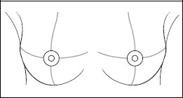
Clinical Findings

Ultrasound
Mammography
MR Mammography
MRM score | Finding | Points |
Shape | round | 0 |
Border | ill-defined | 1 |
CM Distribution | rim sign | 2 |
Initial Signal Intensity Increase | moderate | 1 |
Post-initial Signal Intensity Character | plateau | 1 |
MRI score (points) |
| 5 |
MRI BI-RADS |
| 4 |
 Differential Diagnosis
Right: Carcinoma, adenosis.
BI-RADS Categorization | ||
Clinical Findings | right 1 | left 1 |
Ultrasound | right 3 | left 1 |
Mammography | right 3 | left 1 |
MR Mammography | right 4 | left 1 |
BI-RADS Total | right 4 | left 1 |
Procedure
Histopathological analysis of the lesion in the right breast by US-guided percutaneous core biopsy.
Histopathological analysis of the specimen (right breast)
Chronic fibrocystic mastopathy. No malignancy.
Further procedure
With regard to the discrepancy between the imaging findings in the right breast (in particular MRI) and the histological results, a further percutaneous core biopsy was necessary for increased accuracy. An MR-guided vacuum biopsy was recommended. Instead, in the present case, an excisional biopsy was performed on the lesion in the right breast at the patient’s request.
Histology
Invasive ductal carcinoma measuring 15 mm. Axillary lymph node status normal.
IDC pTIc, pN0(0/17), G2.
Stay updated, free articles. Join our Telegram channel

Full access? Get Clinical Tree


