Case 72
 Clinical Presentation
An 85-year-old woman with recurrent urinary infections.
 Imaging Findings
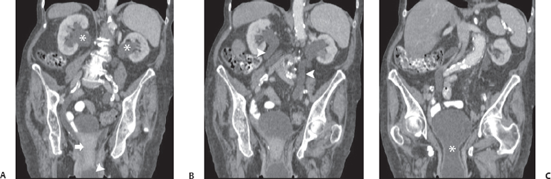
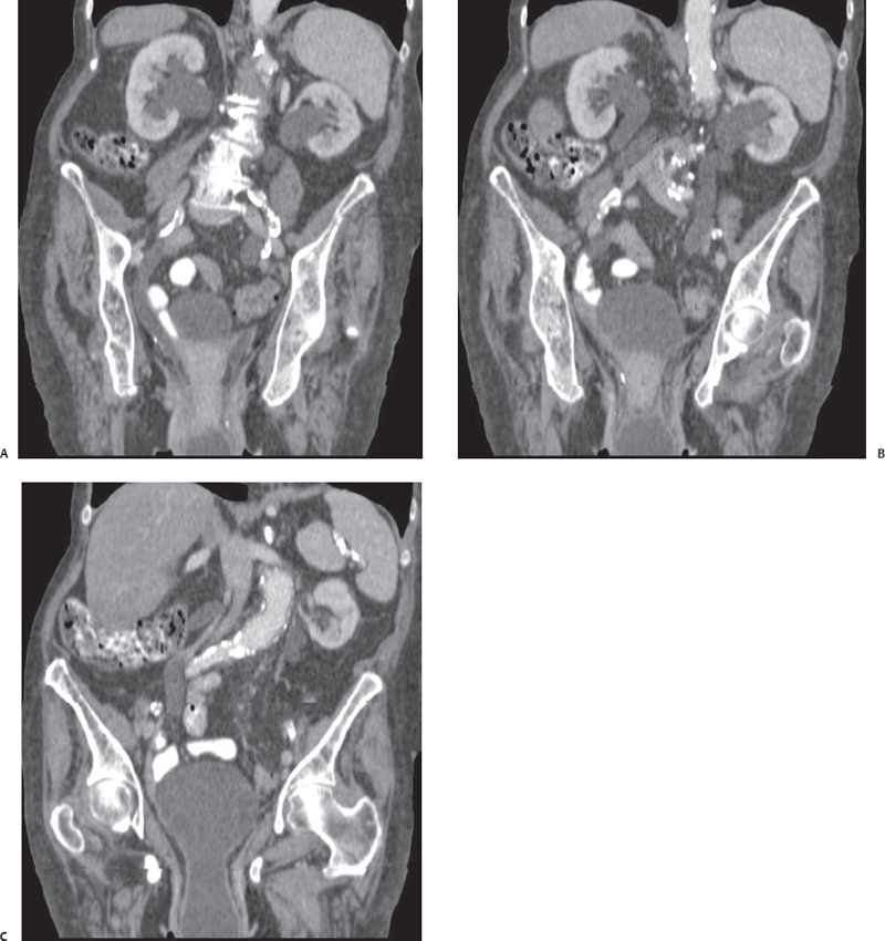
(A) Coronal contrast-enhanced computed tomography (CT) image shows bilateral hydronephrosis (asterisks). In the pelvis, the uterus (arrow) is in an abnormally low location. A cystic structure (arrowhead) is seen in the vagina. (B) Coronal contrast-enhanced CT image anterior to that in Figure A shows the ureters (arrowheads) to be dilated and tortuous, suggesting long-standing obstruction. (C) Coronal contrast-enhanced CT image anterior to that in Figure B shows the cystic structure to be a part of the urinary bladder (asterisk) descending through the pelvic floor.
Stay updated, free articles. Join our Telegram channel

Full access? Get Clinical Tree


