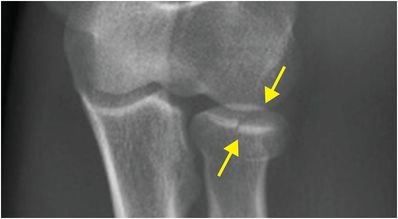
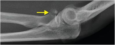
Diagnosis: Radial neck fracture
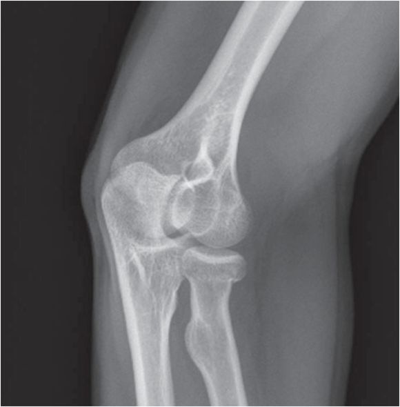
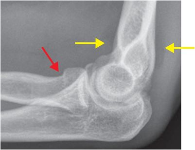
Lateral (left image) and AP radiographs of the elbow demonstrate elevated anterior and posterior fat pads (yellow arrows), indicative of an elbow joint effusion. There is a subtle depressed fracture of the radial neck (red arrows), best appreciated on the AP radiograph.
Discussion
Overview of radial head and neck fractures
Often radiographically occult, fractures through the radial head or neck most commonly occur following a fall onto outstretched hand (FOOSH), with the radial head impacting upon the capitellum.
Radial head or neck fractures can be seen with posterior elbow dislocations and as a component of complex injuries about the elbow.
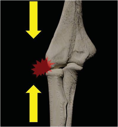
Axial loading at the elbow (yellow arrows), commonly seen in a fall on outstretched hand (FOOSH), causes impact of the radial head on the capitellum with potential for radial head fracture.
Radial head fractures are commonly classified according to the Mason classification system.
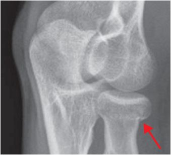
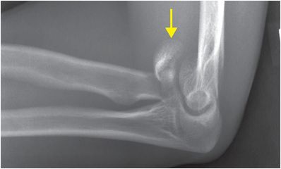
Type I: Minimally displaced (<2 mm displacement) radial head or neck fracture.
AP radiograph of the elbow demonstrates a minimally displaced radial head fracture (arrows), consistent with Mason I fracture.
Type II: Displaced single fracture (>2 mm displacement) with <30% articular involvement.
Lateral radiograph of the elbow demonstrates a displaced fracture of the radial head (arrow) with associated posterior dislocation of the radius at the radiocapitellar articulation. There is also posterior subluxation of the ulna, with perching of the distal humerus on the coronoid process tip.
Type III: Comminuted radial head fracture.
Lateral radiograph of the elbow demonstrates a comminuted radial head fracture (arrow).
Related posts:
 12 68-year-old man with left lower quadrant pain and hypotension
 72 67-year-old female with shoulder pain and limited range of motion following a fall onto an outstretched hand
 35 37-year-old woman with a history of rheumatoid arthritis presenting with non-resolving bilateral effusions and chest pain
 29 19-year-old male presented with acute onset right scrotal pain
 53 42-year-old female presenting with fever and back pain
 66 21-year-old male with quadriplegia after diving into a shallow pond. The patient struck his head against an embankment, with his head flexed, chin against chest
Stay updated, free articles. Join our Telegram channel

Full access? Get Clinical Tree


