Case 73
 Clinical Presentation
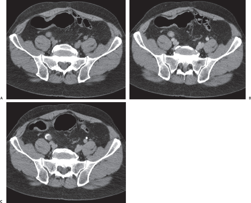
A 68-year-old man with flank pain and hematuria.
 Imaging Findings
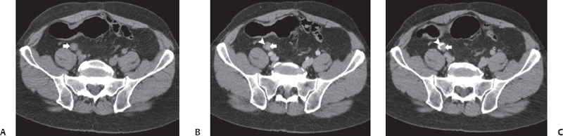
(A) Noncontrast computed tomography (CT) image at the level of the pelvis shows a nodule (arrow) anterior to the right psoas muscle in the expected location of the right ureter. No calcification is seen. (B) Contrast-enhanced CT image at the same level as in Figure A shows enhancement of a portion of the nodule (arrow). Because of the enhancement of the mass, a thin crescent (arrowhead) of fluid density is now visible on one side of the nodule. (C) Excretory phase image at same level as Figures A and B shows opacification of the fluid crescent (arrowhead) by excreted contrast, which outlines the smooth nodule (arrow) and produces a filling defect in the ureter. The left ureter (black asterisk) is normal. No lymphadenopathy is seen.
 Differential Diagnosis
• Ureteric cancer: An enhancing mass in the ureter that obstructs the ureter is characteristic.
• Ureteric obstruction by blood clot: An obstructing blood clot in the ureter may appear as an obstructing lesion with soft-tissue or higher attenuation. However, enhancement is not expected.
• Ureteric stone:
Stay updated, free articles. Join our Telegram channel

Full access? Get Clinical Tree


