CASE 73 A 75-year-old man on anticoagulant treatment presents with easily bleeding gums and abdominal discomfort. Fig. 73.1 Noncontrast axial CT image through the left adrenal gland shows a rounded left adrenal mass (arrow) that is hyperdense on CT with a density of 55 to 60 HU. A small hyperdense lesion is seen in the left adrenal gland (Fig. 73.1). Adrenal hemorrhage Adrenal hemorrhage is a rare condition that usually occurs in response to stressful stimuli, such as infection, septicemia, blunt abdominal trauma, hypotensive shock, burns, neonatal asphyxia, coagulopathies with different causes, and inferior vena caval or renal venous thrombosis. Unilateral right adrenal involvement is most common. Neonates with a history of asphyxia and/or septicemia can present with an abdominal mass, hypotension, hemodynamic shock, anemia, and jaundice and, rarely, with scrotal hematoma. In adults with coagulopathies, blunt trauma, and septicemia, adrenal hemorrhage is detected as an incidental finding on a work-up for internal concealed bleeds.
Clinical Presentation
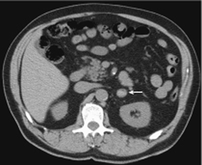
Radiologic Findings
Diagnosis
Differential Diagnosis
Discussion
Background
Clinical Findings
Related posts:


Stay updated, free articles. Join our Telegram channel

Full access? Get Clinical Tree


