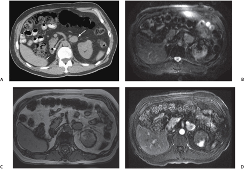
CASE 74 A 70-year-old man with a history of right nephrectomy for renal cell carcinoma comes for restaging of a malignancy. Fig. 74.1 (A) Axial noncontrast CT image in a 70-year-old man with a history of nephrectomy for renal cell carcinoma shows a well-defined, isodense left adrenal mass lesion (arrow). (B) Axial fat-saturated T2-weighted image in the same patient shows the lesion to be hyperintense. (C) On T1-weighted out-of-phase gradient echo imaging, the lesion shows no drop in signal intensity. (D) Axial contrast-enhanced T1-weighted imaging in the same patient shows intense enhancement of the lesion on an early arterial phase scan. Axial computed tomography (CT) and magnetic resonance (MR) images show a left adrenal mass that appears isointense on T1-weighted imaging, moderately hyperintense on T2-weighted imaging, does not show a drop in signal intensity on out-of-phase spoiled gradient echo images, and shows intense enhancement on contrast administration (Fig. 74.1). Metastatic renal cell carcinoma to the left adrenal gland
Clinical Presentation
Radiologic Findings
Diagnosis
Differential Diagnosis
Discussion
Background
Related posts:


Stay updated, free articles. Join our Telegram channel

Full access? Get Clinical Tree


