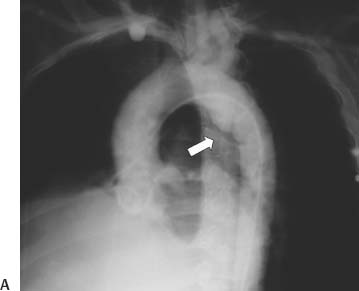
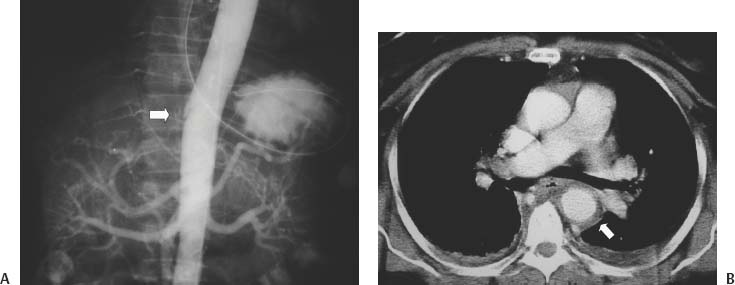
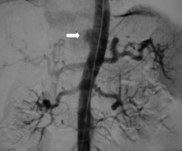
CASE 74 A 35-year-old male presented to the emergency department after a motor vehicle accident, complaining of chest pain and shortness of breath. Figure 74-1 (A) A 35-year-old male complained of chest pain after a motor vehicle accident. Conventional aortogram in the left anterior oblique projection revealed a pseudoaneurysm of the descending aorta at the level of the aortic isthmus, as indicated by a saccular outpouching of the aorta with irregular contour and an intimal flap (arrow). (B) Contrast-enhanced CT confirms pseudoaneurysm related to traumatic aortic injury, separated from the aortic lumen by an intimal flap (arrow). Conventional angiography and contrast-enhanced CT revealed a pseudoaneurysm of the descending aorta at and below the level of the aortic isthmus (Fig. 74-1A). Traumatic aortic injury. The patient underwent left thoracotomy and placement of an interposition graft across the pseudoaneurysm. Contrast material 5F Micropuncture set (Cook, Bloomington, Indiana) 0.035″ standard guidewire (15J; Boston Scientific, Natick, Massachusetts) 5F pigtail flush catheter (Cook, Bloomington, Indiana) Figure 74-3 A 31-year-old male with traumatic aortic injury. Digital subtraction aortogram shows pseudo-aneurysm (arrow) at the level of the diaphragm. Traumatic aortic injury typically occurs during rapid deceleration, most commonly related to motor vehicle accidents. In 80% of cases complete transection occurs, and the victim expires before reaching the hospital. In 20%, partial transection occurs, and the victim survives long enough for evaluation and treatment. Transection occurs at points of aortic fixation within the chest where the shear stresses are greatest. The most common site of injury for survivors is the aortic isthmus, just distal to the takeoff of the subclavian artery (over 90%). Other sites include the aortic isthmus with the proximal ascending aorta, the proximal ascending aorta alone, the distal ascending aorta, and the descending aorta, particularly at the level of the diaphragm (Figs. 74-2A, 74-3
Clinical Presentation
Radiologic Studies
Diagnosis
Treatment
Equipment
Discussion
Background
![]()
Stay updated, free articles. Join our Telegram channel


Full access? Get Clinical Tree


