Diagnosis: Volar plate avulsion fracture
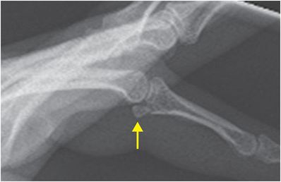
Lateral radiograph of the second digit demonstrates a volar plate avulsion fracture (arrow) at the proximal interphalangeal (PIP) joint, without dislocation.
Discussion
Overview of volar plate injury
The volar plate is a ligamentous structure of the PIP joint that prevents hyperextension.
A volar plate injury may involve the ligament only, or may involve an osseous avulsion fracture. A volar plate avulsion is a fracture of the volar aspect of the articular base of the middle phalanx, and is commonly caused by a hyperextension injury with axial pressure applied to the fingertip.
Volar plate fractures are often associated with dorsal subluxation or dislocation at the PIP joint.
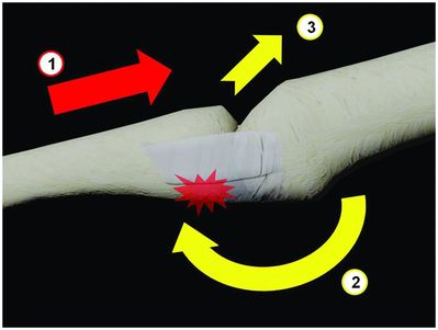
Axial loading along the shaft of the middle phalanx (1) and hyperextension at the PIP joint (2) can cause avulsion injuries at the base of the middle phalanx with dorsal subluxation at the PIP joint (3).
Classification of volar plate fractures
Volar plate fractures are classified according to the stability of the joint after reduction. The volar plate may remain inserted on an avulsed distal fracture fragment, but collateral ligaments may be disrupted, or can remain inserted on either the volar or dorsal fragments.
Stable fractures are generally small fractures with <40% articular surface involvement, as the proper collateral ligaments are not functionally disrupted. This type of stable fracture is often referred to as a volar plate avulsion “chip” injury.
Unstable fractures most often involve >40% of the articular surface, commonly with frank dorsal dislocation. Significant dislocation implies complete functional disruption of the volar plate, with associated collateral ligament disruption or proper collateral ligament insertion on the small volar fracture fragment.
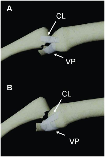
Illustration of the PIP joint in a lateral projection with volar plate (VP) fractures and dorsal phalanx dislocation. Stability is maintained when the fracture fragment is small and the proper collateral ligament (CL) maintains its insertion on the larger dorsal fracture component (A). When the fracture fragment is large, the proper collateral ligament either inserts onto the fracture fragment (as diagrammed in B), or the ligament is frankly disrupted and the fracture is unstable.
Imaging of volar plate injuries
PA and true lateral finger radiographs are essential to diagnosis, though the presence or size of the fracture fragment may not reflect the degree of functional instability. The “V” sign represents divergence of the proximal and middle phalanges at the dorsal aspect of the PIP articulation, as seen on the lateral radiograph. The presence of a “V” sign indicates dorsal subluxation and probable instability.
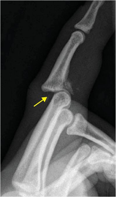
Lateral radiograph of the index finger demonstrates a “V” sign (arrow), with dorsal subluxation of the middle phalanx and a volar plate fracture.
Stay updated, free articles. Join our Telegram channel

Full access? Get Clinical Tree


