Case 75
 Clinical Presentation
A 32-year-old woman with recurrent left pelvic pain.
 Imaging Findings
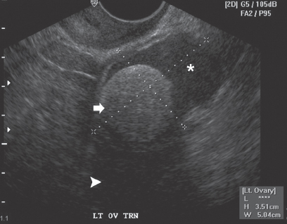
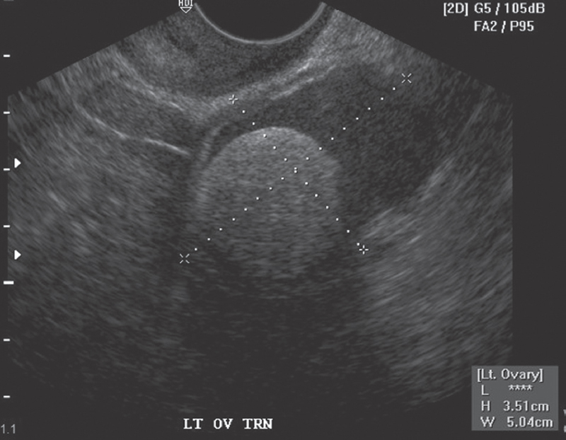
Transvaginal ultrasound image of the left ovary shows a complex ovarian cystic mass with mildly echogenic contents (asterisk). The mass has a large hyperechoic area (arrow) with distal acoustic shadowing (arrowhead). No calcifications are seen.
 Differential Diagnosis
• Ovarian dermoid cyst: A focal noncalcified hyperechoic area with distal acoustic shadowing within a cystic ovarian mass is characteristic.
• Ovarian neoplasm: A malignant ovarian neoplasm can appear as a complex cyst with echogenic soft-tissue areas in the fluid-containing cyst. However, distal acoustic shadowing behind the echogenic area is not expected.
• Hemorrhagic follicle: A blood clot may appear as a solid area floating within a cyst. However, acoustic shadowing is not expected.
Stay updated, free articles. Join our Telegram channel

Full access? Get Clinical Tree


